Review
. 2011 Apr 15;508(2):123-37.
doi: 10.1016/j.abb.2010.12.019. Epub 2010 Dec 19.

Keratin gene mutations in disorders of human skin and its appendages

Affiliations
Review

Keratin gene mutations in disorders of human skin and its appendages

Jean Christopher Chamcheu et al. Arch Biochem Biophys. .

Abstract

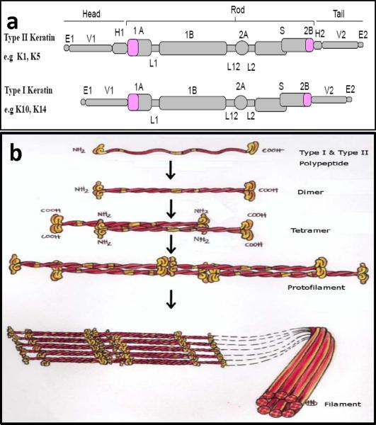
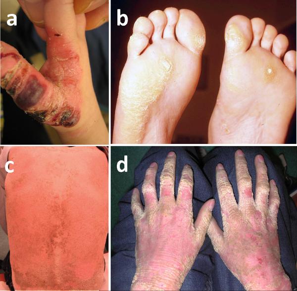
Keratins, the major structural protein of all epithelia are a diverse group of cytoskeletal scaffolding proteins that form intermediate filament networks, providing structural support to keratinocytes that maintain the integrity of the skin. Expression of keratin genes is usually regulated by differentiation of the epidermal cells within the stratifying squamous epithelium. Amongst the 54 known functional keratin genes in humans, about 22 different genes including, the cornea, hair and hair follicle-specific keratins have been implicated in a wide range of hereditary diseases. The exact phenotype of each disease usually reflects the spatial expression level and the types of mutated keratin genes, the location of the mutations and their consequences at sub-cellular levels as well as other epigenetic and/or environmental factors. The identification of specific pathogenic mutations in keratin disorders formed the basis of our understanding that led to re-classification, improved diagnosis with prognostic implications, prenatal testing and genetic counseling in severe keratin genodermatoses. Molecular defects in cutaneous keratin genes encoding for keratin intermediate filaments (KIFs) causes keratinocytes and tissue-specific fragility, accounting for a large number of genetic disorders in human skin and its appendages. These diseases are characterized by keratinocytes fragility (cytolysis), intra-epidermal blistering, hyperkeratosis, and keratin filament aggregation in severely affected tissues. Examples include epidermolysis bullosa simplex (EBS; K5, K14), keratinopathic ichthyosis (KPI; K1, K2, K10) i.e. epidermolytic ichthyosis (EI; K1, K10) and ichthyosis bullosa of Siemens (IBS; K2), pachyonychia congenita (PC; K6a, K6b, K16, K17), epidermolytic palmo-plantar keratoderma (EPPK; K9, (K1)), monilethrix (K81, K83, K86), ectodermal dysplasia (ED; K85) and steatocystoma multiplex. These keratins also have been identified to have roles in apoptosis, cell proliferation, wound healing, tissue polarity and remodeling. This review summarizes and discusses the clinical, ultrastructural, molecular genetics and biochemical characteristics of a broad spectrum of keratin-related genodermatoses, with special clinical emphasis on EBS, EI and PC. We also highlight current and emerging model tools for prognostic future therapies. Hopefully, disease modeling and in-depth understanding of the molecular pathogenesis of the diseases may lead to the development of novel therapies for several hereditary cutaneous diseases.


Figures

Figure 1
Figure 1. Skin structure and keratin intermediate filament in triton extracted cultured keratinocyte cytoskeleton
(a) Cross section of the human skin tissue showing the three distinct skin components, (b) Triton X-100 extracted keratinocyte cytoskeleton in culture stained with keratin 14 antibody (LL002; green filaments) shows a dense network of keratin intermediate filament bundles. The protein scaffold linked to its associated complexes forms the main resilience structure of the epithelial keratinocytes. (Reproduced from [133] with permission from the publisher)
Figure 2
Figure 2. Molecular structure and assembly of keratin intermediate filaments (KIF)
(a) Schematic representation of type I and type II keratin polypeptide domain structural organization. Of the 54 different human keratin genes, each keratin molecule consists of a central alpha helical rod domain which is composed of four helical segments, 1A, IB, 2A and 2B that are interrupted by three flexible non-helical linker domains L1, L12 and L2. The rod domain begins and ends with highly conserved sequence motifs, helix initiation (HIP) and helix termination (HTP) peptides and is flanked by head and tail domains, respectively, (b) Keratin intermediate filaments assembly; Keratin polymerization obligatorily begins with the formation of coiled-coil obligate heterodimer structures involving winding around each other of the central rod domains of type I and type II polypeptides, a requirement underlying the pair wise transcriptional regulation of keratin genes in vivo. The heterodimers then associate (side-by-side) and assemble in an overlapping staggered and antiparallel fashion to form stable tetramers. Tetramers then associate end-to-end to form protofilaments and finally, four protofibrils laterally build keratin intermediate filaments. Each filament contains approximately eight protofilaments wound around each other in a rope-like structure, forming the 10–12 nm wide KIF network. (Reproduced from [133] with permission from the publisher)
Figure 3
Figure 3. Clinical features of epidermolysis bullosa simplex (EBS) and epidermolytic ichthyosis (EI)
(a) A child's hand with severe generalized Dowling-Meara form of EBS, characterized by widespread herpetiform blister that heals without scar formation, (b) An Adult with severe EBS-DM whose feet present with painful plantar callosities, (c) Epidermolytic ichthyosis patient showing sharp massive hyperkeratosis of the lower back and (d) a diffuse hyperkeratosis of the hand and flexures with an erythromatous background. (Reproduced from [133] with permission from the publisher)
Figure 4
Figure 4. Triton extracted monolayer cultures of keratinocytes cytoskeleton and 3-D epidermis engineered with keratinocytes harboring mutations
Heat stress of keratin cytoskeleton reveals pathognomonic keratin-aggregates in keratinocytes harboring (a) K5_p.475Gly>Glu mutation in severe generalized EBS-DM keratinocyte cell line (EB11) when stained with keratin 5 antibody, (b) K10_p.l56Arg>Gly mutation in a moderate EI-phenotype keratinocyte cell line (EH21) when stained with keratin 10 antibody. Keratin 10 expression was induced in submerged culture by calcium induced differentiation of keratinocytes. (c) Haematoxylin staining of organotypic epidermis generated using immortalized epidermolytic ichthyosis (EI: EH31) keratinocyte cell lines derived from a severely affected patient's organotypic epidermis and without heat-stress. The reconstructed epidermis here shows well-defined basal layer, superficial cleft or cytolysis at the suprabasal layers (arrows) leaving unperturbed basal and cornified layers, a histological feature that mimics the in vivo phenotype of the patient, (d) Haematoxylin staining of organotypic epidermis generated using immortalized epidermolysis bullosa simplex (EB11) keratinocyte cell lines derived from a severely affected EBS-DM patient's organotypic epidermis and after heat-stress. The organotypic epidermis shows well defined basal layer with some basal cell cytolysis or cleft formation reminiscent of in vivo severe generalized EBS phenotype. All organotypic epidermis was harvested after 12 day post-lifted at air-liquid interface.

References

    1. Tabolli S, Pagliarello C, Uras C, Di Pietro C, Zambruno G, Castiglia D, Sampogna F, Abeni D. Family burden in epidermolysis bullosa is high independent of disease type/subtype. Acta Derm Venereol. 2010;90:607–611. - PubMed
    1. Pagliarello C, Tabolli S. Factors affecting quality of life in epidermolysis bullosa. Expert Rev Pharmacoecon Outcomes Res. 2010;10:329–338. - PubMed
    1. Coulombe PA, Hutton ME, Vassar R, Fuchs E. A function for keratins and a common thread among different types of epidermolysis bullosa simplex diseases. J Cell Biol. 1991;115:1661–1674. - PMC - PubMed
    1. Gu X, Xu F, Wang X, Gao X, Zhao Q. Molecular cloning and expression of a novel CYP26 gene (cyp26d1) during zebrafish early development. Gene Expr Patterns. 2005;5:733–739. - PubMed
    1. Moll R, Franke WW, Volc-Platzer B, Krepler R. Different keratin polypeptides in epidermis and other epithelia of human skin: a specific cytokeratin of molecular weight 46,000 in epithelia of the pilosebaceous tract and basal cell epitheliomas. J Cell Biol. 1982;95:285–295. - PMC - PubMed

Publication types