. 2011 Jul;25(5):1025-35.
doi: 10.1016/j.bbi.2010.12.008. Epub 2010 Dec 19.

Neuroinflammation modulates distinct regional and temporal clinical responses in ALS mice

Affiliations

Neuroinflammation modulates distinct regional and temporal clinical responses in ALS mice

David R Beers et al. Brain Behav Immun. 2011 Jul.

Abstract

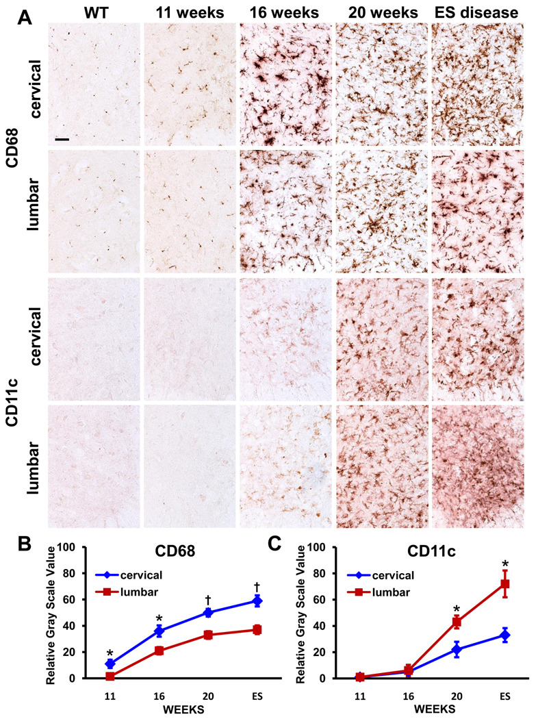
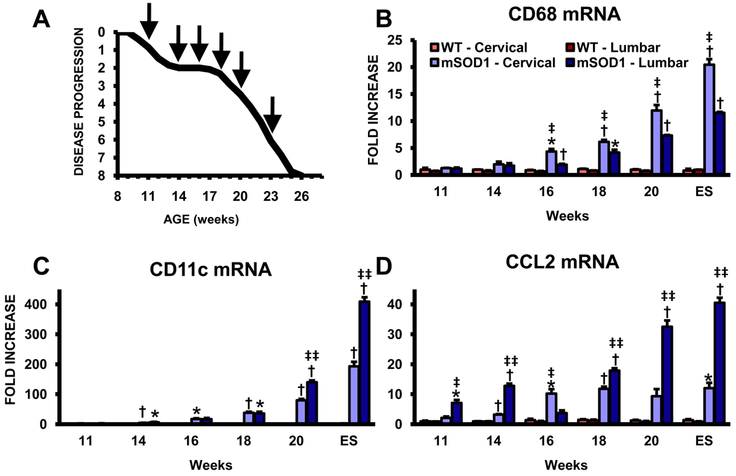
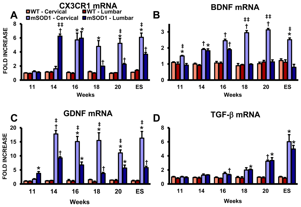
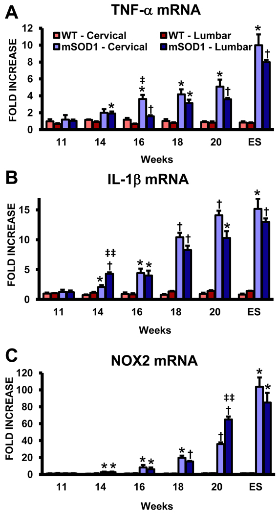
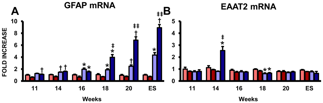
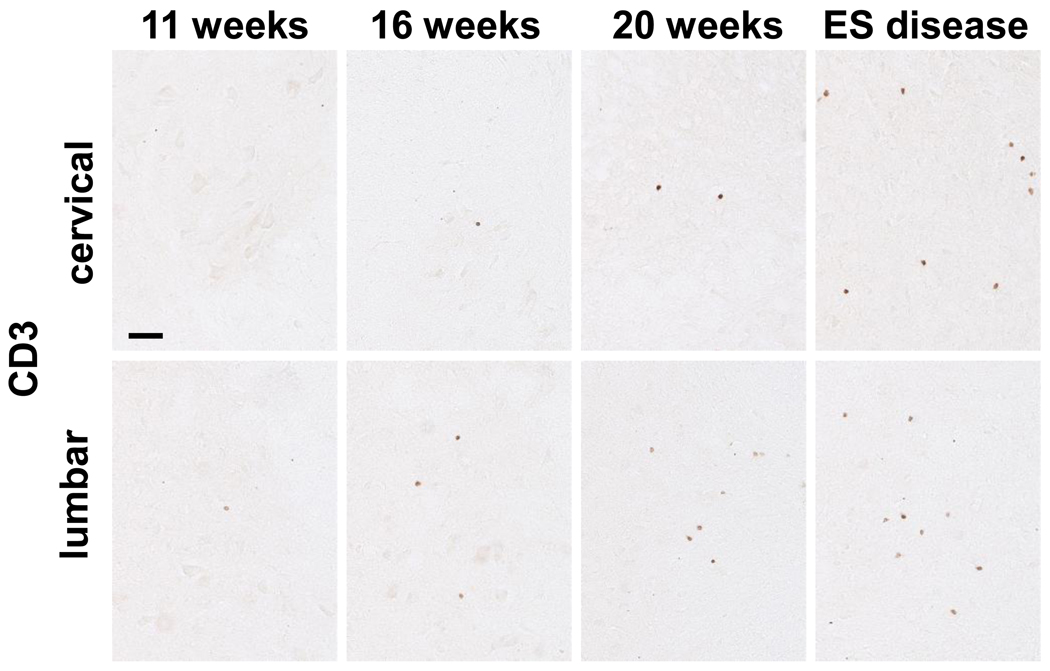
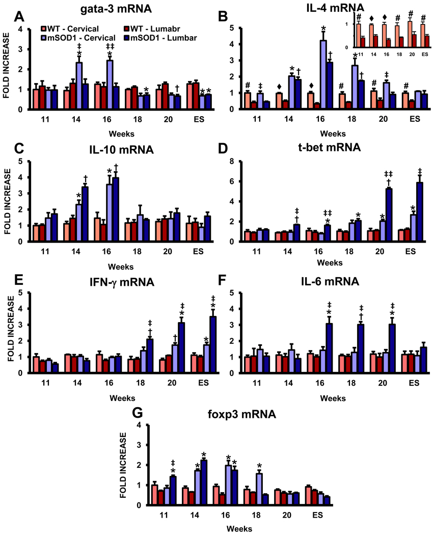
An inflammatory response is a pathological hallmark of amyotrophic lateral sclerosis (ALS), a relentless and devastating degenerative disease of motoneurons. This response is not simply a late consequence of motoneuron degeneration, but actively contributes to the balance between neuroprotection and neurotoxicity; initially infiltrating lymphocytes and microglia slow disease progression, while later, they contribute to the acceleration of disease. Since motor weakness begins in the hindlimbs of ALS mice and only later involves the forelimbs, we determined whether differential protective versus injurious inflammatory responses in the cervical and lumbar spinal cords explained the temporally distinct clinical disease courses between the limbs of these mice. Densitometric evaluation of immunohistochemical sections and quantitative RT-PCR (qRT-PCR) demonstrated that CD68 and CD11c were differentially increased in their spinals cords. qRT-PCR revealed that protective and anti-inflammatory factors, including BDNF, GDNF, and IL-4, were increased in the cervical region compared with the lumbar region. In contrast, the toxic markers TNF-α, IL-1β and NOX2 were not different between ALS mice cervical and lumbar regions. T lymphocytes were observed infiltrating lumbar spinal cords of ALS mice prior to the cervical region; mRNA levels of the transcription factor gata-3 (Th2 response) were differentially elevated in the cervical cord of ALS mice whereas t-bet (Th1 response) was increased in the lumbar cord. These results reinforce the important balance between specific protective/injurious inflammatory immune responses in modulating clinical outcomes and suggest that the delayed forelimb motor weakness in ALS mice is partially explained by augmented protective responses in the cervical spinal cords.


Conflict of interest statement

Conflict of interest statement

All authors declare that there are no conflicts of interest.

Figures

Fig. 1
Fig. 1
Immunohistochemical evaluations of CD68 and CD11c in the cervical and lumbar spinal cords of ALS mice. (A) CD68 is relatively increased in the cervical region whereas CD11c is relatively increased in lumbar region of these mice. (B) and (C) Densitometric analyses confirm the relative difference of CD68 and CD11c immuno-signals. * ≤ 0.05, ALS mice cervical region compared with their lumbar region; † ≤ 0.01, ALS mice cervical region compared with their lumbar region. Scale bar: 50 µm.
Fig. 2
Fig. 2
Quantitative RT-PCR analyses reveal differentially elevated markers for phagocytic and dendritic cell phenotypes in ALS mice. (A) Graph of the disease progression curve and time points (arrows) when qRT-PCR assays were performed on ALS mice; based upon the BASH score. Each time point represents the mean of n = 3 mice. Notice the stable disease phase from 14 to 18 weeks and the rapidly progressing phase subsequent to 18 weeks. (B) mRNA levels for CD68, a LAMP protein expressed on phagocytic cells CD68. (C) CD11c mRNA levels. Notice the scale of the ordinate in this graph. (D) CCL2 mRNA levels. * ≤ 0.05, ALS mice spinal cord regions compared with the same regions from WT mice; † ≤ 0.01, ALS mice spinal cord regions compared with the same regions from WT mice; ‡ ≤ 0.05, ALS mice cervical spinal cords compared with ALS mice lumbar spinal cords, ‡‡ ≤ 0.01, ALS mice cervical spinal cords compared with ALS mice lumbar spinal cords.
Fig. 3
Fig. 3
Anti-inflammatory and neurotrophic factors are increased in the cervical cords of ALS mice. (A) CX3CR1 (fractalkine receptor) mRNA levels. (B) mRNA levels for BDNF. (C) GDNF mRNA levels. (D) The mRNA levels of TGF-β. Each time point represents the mean of n = 3 mice. * ≤ 0.05, ALS mice spinal cord regions compared with the same regions from WT mice; † ≤ 0.01, ALS mice spinal cord regions compared with the same regions from WT mice; ‡ ≤ 0.05, ALS mice cervical spinal cords compared with ALS mice lumbar spinal cords, ‡‡ ≤ 0.01, ALS mice cervical spinal cords compared with ALS mice lumbar spinal cords.
Fig. 4
Fig. 4
An equivalent up-regulation of mRNA for toxic factors in the cervical and lumbar spinal cords of ALS mice. (A) The mRNA for TNF-α. (B) IL-1β mRNA levels. (C) NOX2 mRNA levels. Notice the scale of the ordinate in this graph. Each time point represents the mean of n = 3 mice. * ≤ 0.05, ALS mice spinal cord regions compared with the same regions from WT mice; † ≤ 0.01, ALS mice spinal cord regions compared with the same regions from WT mice; ‡ ≤ 0.05, ALS mice cervical spinal cords compared with ALS mice lumbar spinal cords, ‡‡ ≤ 0.01, ALS mice cervical spinal cords compared with ALS mice lumbar spinal cords.
Fig. 5
Fig. 5
Expression of astrocyte mRNAs in the cervical and lumbar spinal cords of ALS mice. (A) The mRNA for GFAP, a marker of astrocytes and astrocytosis. (B) The expression of EAAT2, the astroglial glutamate transporter responsible for the removal of glutamate from the synaptic cleft. Each time point represents the mean of n = 3 mice. * ≤ 0.05, ALS mice spinal cord regions compared with the same regions from WT mice; † ≤ 0.01, ALS mice spinal cord regions compared with the same regions from WT mice; ‡ ≤ 0.05, ALS mice cervical spinal cords compared with ALS mice lumbar spinal cords, ‡‡ ≤ 0.01, ALS mice cervical spinal cords compared with ALS mice lumbar spinal cords.
Fig. 6
Fig. 6
Immunohistochemical evaluations of CD3 in the cervical and lumbar spinal cords of ALS mice. Scale bar: 50 µm.
Fig. 7
Fig. 7
Expression of lymphocyte mRNAs is elevated in the lumbar spinal cords of ALS mice. (A) mRNA for gata-3, a master transcription factor preferentially expressed in Th2 lymphocytes. (B) IL-4 mRNA levels, the prototypic anti-inflammatory cytokine released by Th2 lymphocytes. There was 2–2.5 fold more in the cervical cords of WT mice compared with their lumbar regions at all time points assayed (insert); IL-4 was the only factor assayed that was different in the two spinal cord regions of WT mice. (C) IL-10 mRNA expression levels. (D) Expression levels of t-bet mRNA, a master transcription factor for Th1 lymphocytes. (E) The mRNA for IFN-γ, the prototypic pro-inflammatory cytokine released by Th1 lymphocytes. (F) The expression level for IL-6, another Th1 induced cytokine. (G) mRNA levels for foxp3, a transcription factor currently accepted as a reliable marker of Tregs. Each time point represents the mean of n = 3 mice. * ≤ 0.05, ALS mice spinal cord regions compared with the same regions from WT mice; † ≤ 0.01, ALS mice spinal cord regions compared with the same regions from WT mice; ‡ ≤ 0.05, ALS mice cervical spinal cords compared with ALS mice lumbar spinal cords, ‡‡ ≤ 0.01, ALS mice cervical spinal cords compared with ALS mice lumbar spinal cords; # ≤ 0.05, WT mice cervical spinal cords compared with WT mice lumbar spinal cords; ♦ ≤ 0.01, WT mice cervical spinal cords compared with WT mice lumbar spinal cords.

References

    1. Ajami B, Bennett JL, Krieger C, Tetzlaff W, Rossi FMV. Local self-renewal can sustain CNS microglia maintenance and function throughout adult life. Nat. Neurosci. 2007;10:1538–1543. - PubMed
    1. Appel SH, Beers DR, Henkel JS. T cell-microglial dialogue in Parkinson's disease and amyotrophic lateral sclerosis: are we listening? Trends Immunol. 2010;31:7–17. - PMC - PubMed
    1. Arvin KL, Han BH, Du Y, Lin SZ, Paul SM, Holtzman DM. Minocycline markedly protects the neonatal brain against hypoxic-ischemic injury. Ann Neurol. 2002;52:54–61. - PubMed
    1. Bachoo RM, Kim RS, Ligon KL, Maher EA, Brennan C, Billings N, Chan S, Li C, Rowitch DH, Wong WH, DePinho RA. Molecular diversity of astrocytes with implications for neurological disorders. Proc. Natl. Acad. Sci. USA. 2004;101:8384–8389. - PMC - PubMed
    1. Banerjee R, Mosley RL, Reynolds AD, Dhar A, Jackson-Lewis V, Gordon PH, Przedborski S, Gendelman HE. Adaptive immune neuroprotection in G93A-SOD1 amyotrophic lateral sclerosis mice. PLoS ONE. 2008;3:e2740. - PMC - PubMed

Publication types

MeSH terms