Review
. 2011 Feb;21(1):128-36.
doi: 10.1016/j.sbi.2010.11.006. Epub 2010 Dec 19.

Nucleosome structural studies

Affiliations
Review

Nucleosome structural studies

Song Tan et al. Curr Opin Struct Biol. 2011 Feb.

Abstract

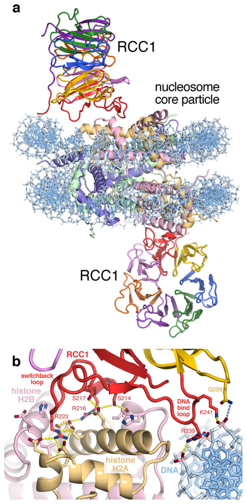
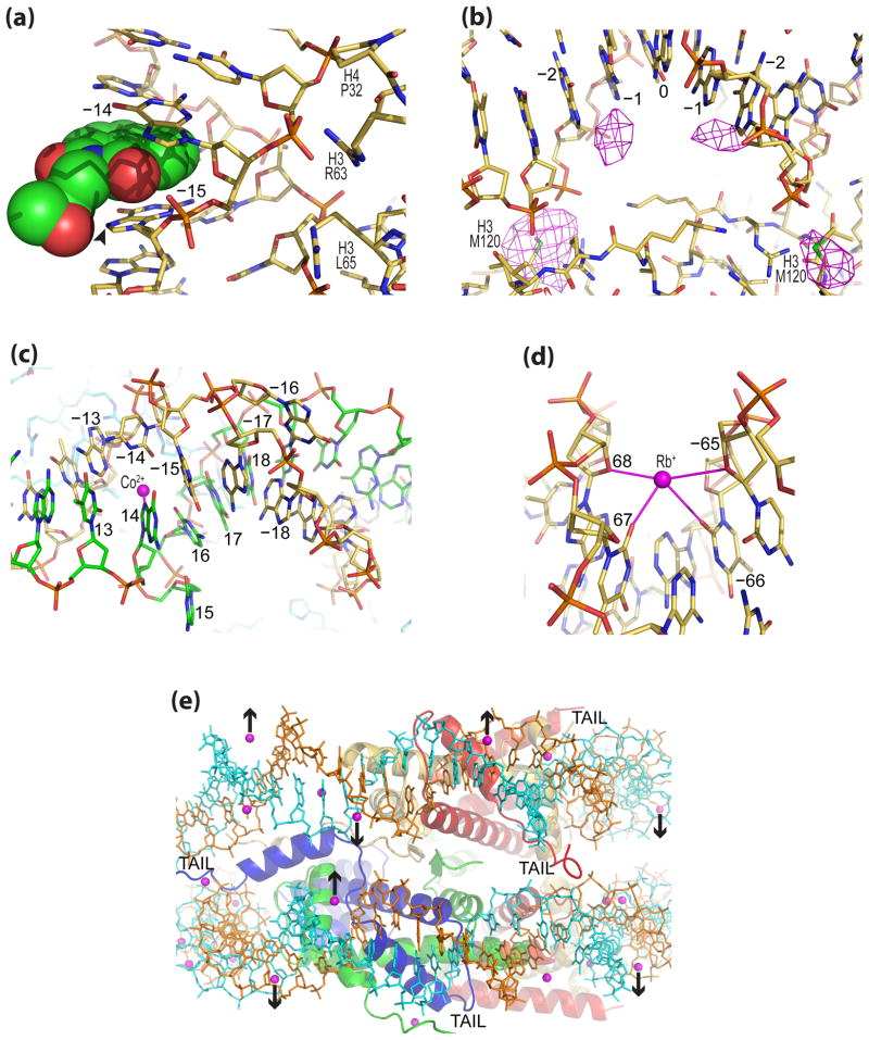
Chromatin plays a fundamental role in eukaryotic genomic regulation, and the increasing awareness of the importance of epigenetic processes in human health and disease emphasizes the need for understanding the structure and function of the nucleosome. Recent advances in chromatin structural studies, including the first structures of nucleosomes containing the Widom 601 sequence and the structure of a chromatin protein-nucleosome assembly, have provided new insight into stretching of nucleosomal DNA, nucleosome positioning, binding of metal ions, drugs and therapeutic candidates to nucleosomes, and nucleosome recognition by nuclear proteins. These discoveries ensure promising future prospects for unravelling structural attributes of chromatin.


Conflict of interest statement

Conflict of interest statement: The authors declare that they have no conflict of interest.

Figures

Figure 1
Figure 1
Crystal structure of the RCC1-nucleosome complex (PDB: 3MVD). (a) Overview of RCC1-nucleosome complex shows that RCC1 uses loops and its N-terminal arm to engage the nucleosome. (b) RCC1 uses its switchback loop to bind to the histone dimer acidic patch and an adjacent DNA-binding loop to interact with nucleosomal DNA.
Figure 2
Figure 2
DNA binding and stretching in the nucleosome core. (a) Comparison of DNA sequence and histone-DNA register in NCP crystal structures (PDB: 1KX5, 2NZD, 1KX3, 2CV5, 1KX4, 3LEL, 3LZ0/3LZ1, 3MVD, top to bottom). All particles are with Xenopus laevis histones, with the exception of hNCP146, which contains histones from Homo sapiens. Asterisks denote severe kinks associated with regions of stretching (underlined in magenta). (b) View of the NCP147 crystal structure down the DNA superhelix axis for approximately one half of the particle (PDB: 1KX5). Numbers correspond to SHL, and alphanumeric entities refer to the DNA-binding histone motifs (α1, alpha helix 1; L1, loop 1; L2, loop 2). Long magenta arrows indicate potential regions of DNA stretching around SHL ±2 and ±5. Histone proteins are colored blue for H3, green for H4, gold for H2A, and red for H2B. (c) The special histone binding site at SHL ±1.5, which selects for or imposes an extremely narrow minor groove via the sugar clamp motif (space filling; NCP-TA; PDB: 3LEL).
Figure 3
Figure 3
Crystallographic studies of therapeutic candidate, drug and metal cation association with the NCP. (a) Facilitated intercalation of an alkylating antitumor compound (space-filling) between guanine bases at the site of stretch-associated extreme minor groove kinking at SHL ±1.5 in NCP145 creates a hotspot for DNA alkylation by the epoxide group (arrowhead; PDB: 3KUY). (b) Anticancer drug cisplatin adduct formation observed around the nucleosome center in NCP147 (PDB: 3B6F). An anomalous difference map (3.4σ, magenta) shows platinum atom locations between purine bases and at methionine sulfur groups. (c) Co2+ binding (magenta) promotes expulsion of two bases ( 16 and 15) from the double helical stack at SHL 1.5 in NCP147 (PDB: 3MGP). (d) A narrow minor groove favors monovalent cation (Rb+) coordination (magenta) at AT dinucleotide elements in NCP147 (PDB: 3MGR). (e) View along the nucleosomal DNA supergroove of NCP147 showing that minor groove-divalent metal cation (magenta spheres) hydrate binding can influence DNA wrapping (inward-pointing arrows) and nucleosome-nucleosome interactions (outward-pointing arrows; PDB: 3LJA).

References

    1. Khorasanizadeh S. The nucleosome: from genomic organization to genomic regulation. Cell. 2004;116:259–272. - PubMed
    1. Segal E, Widom J. What controls nucleosome positions? Trends Genet. 2009;25 :335–343. - PMC - PubMed
    1. Cairns BR. The logic of chromatin architecture and remodelling at promoters. Nature. 2009;461:193–198. - PubMed
    1. Jiang C, Pugh BF. Nucleosome positioning and gene regulation: advances through genomics. Nat Rev Genet. 2009;10:161–172. - PMC - PubMed
    1. Li B, Carey M, Workman JL. The role of chromatin during transcription. Cell. 2007;128:707–719. - PubMed

Publication types

LinkOut - more resources