

High-resolution functional magnetic resonance imaging mapping of noxious heat and tactile activations along the central sulcus in New World monkeys
- PMID: 21177033
- PMCID: PMC3039029
- DOI: 10.1016/j.pain.2010.10.048
High-resolution functional magnetic resonance imaging mapping of noxious heat and tactile activations along the central sulcus in New World monkeys
Abstract
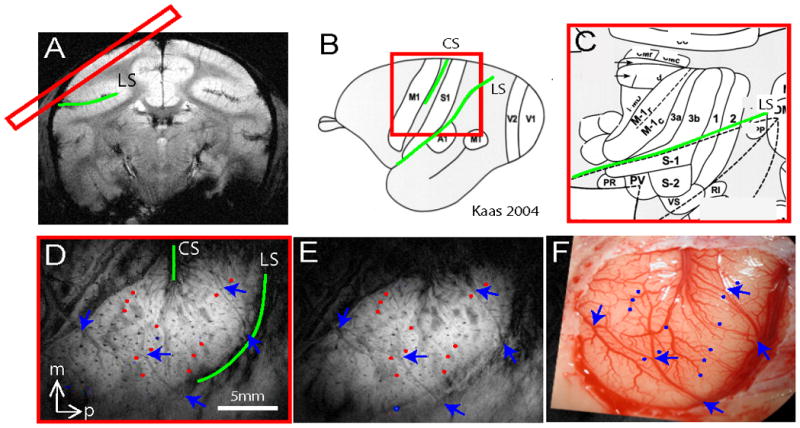
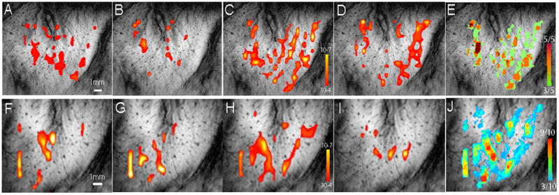
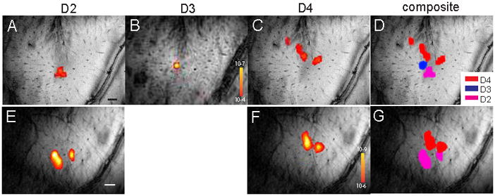
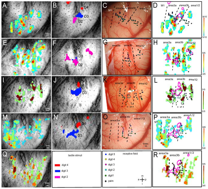
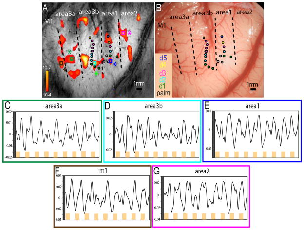
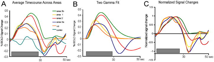
This study mapped the fine-scale functional representation of tactile and noxious heat stimuli in cortical areas around the central sulcus of anesthetized squirrel monkeys by using high-resolution blood oxygen level-dependent (BOLD) fMRI at 9.4T. Noxious heat (47.5°C) stimulation of digits evoked multiple spatially distinct and focal BOLD activations. Consistent activations were observed in areas 3a, 3b, 1, and 2, whereas less frequent activation was present in M1. Compared with tactile activations, thermal nociceptive activations covered more area and formed multiple foci within each functional area. In general, noxious heat activations in area 3b did not colocalize with tactile responses. The spatial relationships of heat and tactile activations in areas 3a and 1/2 varied across animals. Subsequent electrophysiological mapping confirmed that the evoked heat and tactile BOLD signals were somatotopically appropriate. The magnitude and temporal profiles of the BOLD signals to noxious heat stimuli differed across cortical areas. Comparatively late-peaking but stronger signals were observed in areas 3b and 2, whereas earlier-peaking but weaker signals were observed in areas 3a, 1, and M1. In sum, this study not only confirmed the involvement of somatosensory areas of 3a, 3b, and 1, but also identified the engagements of area 2 and M1 in the processing of heat nociceptive inputs. Differential BOLD response profiles of the individual cortical areas along the central sulcus suggest that these areas play different roles in the encoding of nociceptive inputs. Thermal nociceptive and tactile inputs may be processed by different clusters of neurons in different areas. To critically bridge animal and human pain studies, human fMRI was related to primate fMRI and electrophysiology of nociceptive processing, examining the functional role of the primary somatosensory cortex in heat nociception and demonstrating that subregion areas 3a, 3b, 1, 2, and M1 are responsive to noxious heat stimuli.
Copyright © 2010 International Association for the Study of Pain. Published by Elsevier B.V. All rights reserved.
Conflict of interest statement
Figures


Comment in
-
Spatial and temporal patterns of cortical activation underlying pain and tactile sensation.Pain. 2011 Mar;152(3):473-474. doi: 10.1016/j.pain.2010.12.008. Epub 2011 Jan 5. Pain. 2011. PMID: 21211908 No abstract available.
References
-
- Alappat JJ. Motor cortex stimulation for chronic pain: systematic review and meta-analysis of the literature. Neurology. 2009;72(6):577. author reply 77. - PubMed
-
- Apkarian AV, Bushnell MC, Treede RD, Zubieta JK. Human brain mechanisms of pain perception and regulation in health and disease. Eur J Pain. 2005;9(4):463–84. - PubMed
-
- Casey KL, Minoshima S, Morrow TJ, Koeppe RA. Comparison of human cerebral activation pattern during cutaneous warmth, heat pain, and deep cold pain. J Neurophysiol. 1996;76(1):571–81. - PubMed
Publication types
MeSH terms
Substances
Grants and funding
LinkOut - more resources
Full Text Sources
