. 2011 Feb 15;71(4):1374-84.
doi: 10.1158/0008-5472.CAN-10-2238. Epub 2010 Dec 21.

Therapeutic antibody targeting of CD47 eliminates human acute lymphoblastic leukemia

Affiliations

Therapeutic antibody targeting of CD47 eliminates human acute lymphoblastic leukemia

Mark P Chao et al. Cancer Res. .

Abstract

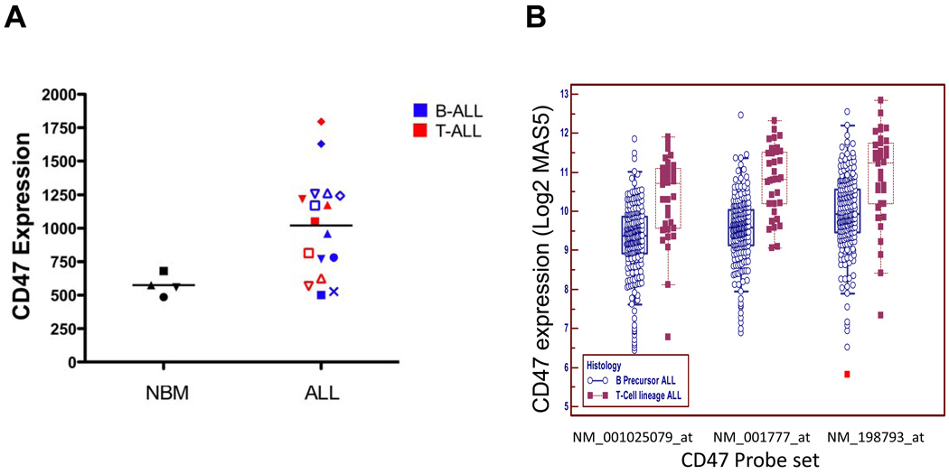
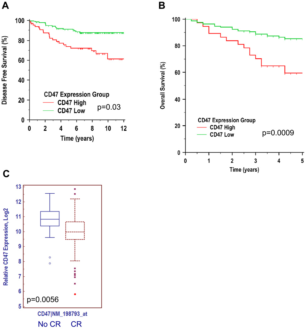
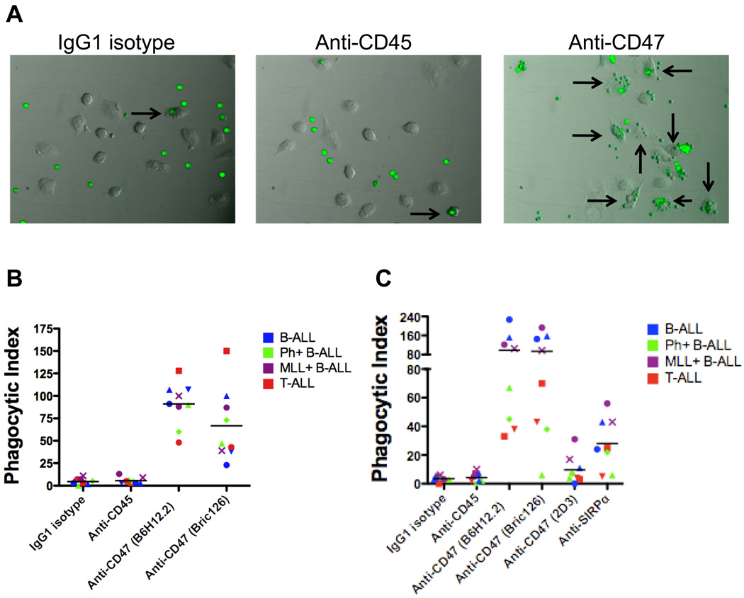
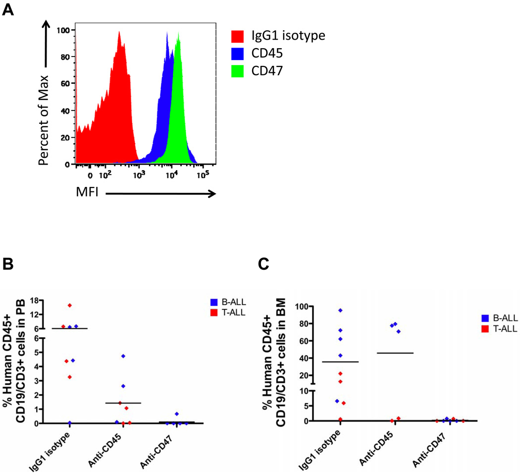
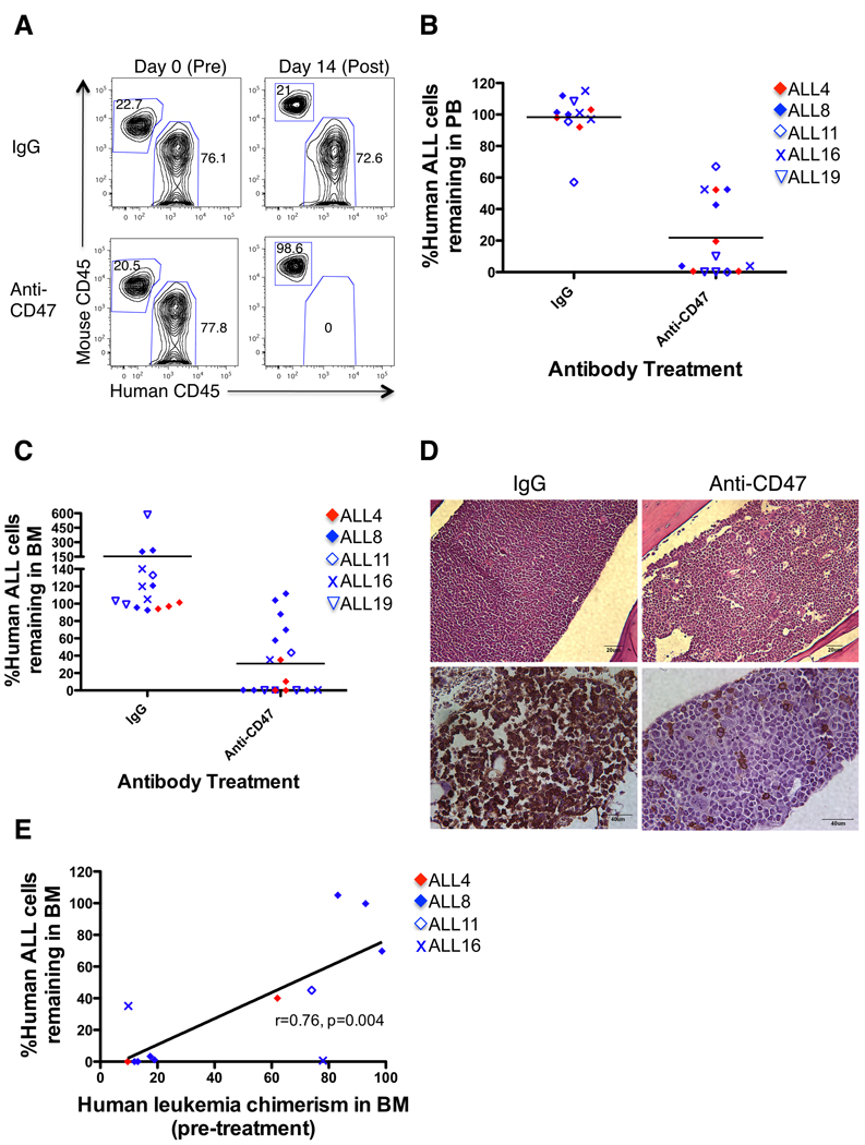
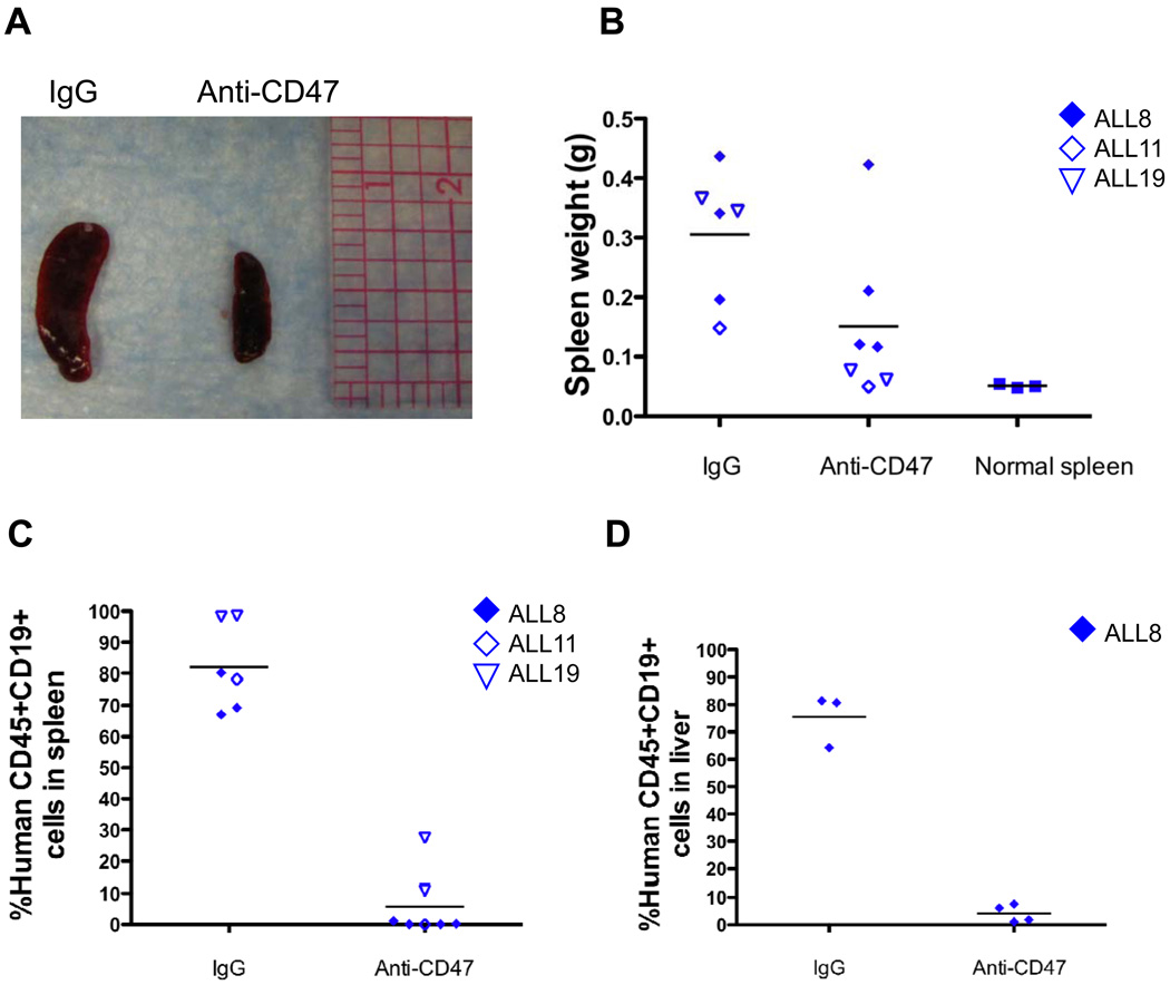
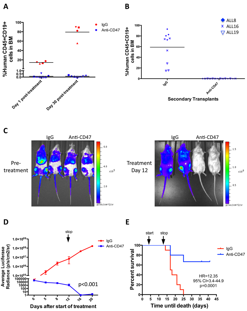
Acute lymphoblastic leukemia (ALL) is the most common pediatric malignancy and constitutes 15% of adult leukemias. Although overall prognosis for pediatric ALL is favorable, high-risk pediatric patients and most adult patients have significantly worse outcomes. Multiagent chemotherapy is standard of care for both pediatric and adult ALL, but is associated with systemic toxicity and long-term side effects and is relatively ineffective against certain ALL subtypes. Recent efforts have focused on the development of targeted therapies for ALL including monoclonal antibodies. Here, we report the identification of CD47, a protein that inhibits phagocytosis, as an antibody target in standard and high-risk ALL. CD47 was found to be more highly expressed on a subset of human ALL patient samples compared with normal cell counterparts and to be an independent predictor of survival and disease refractoriness in several ALL patient cohorts. In addition, a blocking monoclonal antibody against CD47 enabled phagocytosis of ALL cells by macrophages in vitro and inhibited tumor engraftment in vivo. Significantly, anti-CD47 antibody eliminated ALL in the peripheral blood, bone marrow, spleen, and liver of mice engrafted with primary human ALL. These data provide preclinical support for the development of an anti-CD47 antibody therapy for treatment of human ALL.


Figures

Figure 1
Figure 1. CD47 expression is increased on a subset of human ALL cells compared to normal bone marrow
(A) Relative CD47 protein expression was determined on normal human BM cells and human ALL cells (Supplementary Table S1) by flow cytometry. CD47 expression was increased on bulk ALL cells compared to normal BM cells (p=0.006). Within all samples, normalized mean expression (and range) was determined as follows: NBM 575.3 (486.6–680.9), bulk ALL 1020.2 (501.3 – 1794.5), B-ALL 980.5 (501.3 – 1628.4), and T-ALL 1034.9 (626.2 – 1794.5). Differences between mean expression of NBM and T-ALL (p=0.03), NBM and B-ALL (p=0.006) were statistically significant. (B) Relative CD47 mRNA expression was determined in a large cohort of ALL patients (n=254) from (14) and subdivided into B or T-ALL subtypes. Using three different probe sets, CD47 expression was higher in T-ALL patients compared to B-ALL patients (p<0.0001 for all three probe sets).
Figure 2
Figure 2. CD47 expression is an independent prognostic predictor in mixed and high-risk ALL
(A) Pediatric ALL patients (n=360) with mixed risk and treatment (15) were stratified into CD47 high- and low-expressing groups based on an optimal cut point. Disease-free survival (DFS) was determined by Kaplan-Meier analysis (see Supplementary Table S2A). (B) Pediatric ALL patients (n=207) with high-risk (defined by age>10 years, presenting WBC count>50,000/µl, hypodiploidy, and BCR-ABL positive disease) and uniform treatment (18) were stratified into CD47 high- and low-expressing groups using a similar approach as in A (see Supplementary Table S2B). (C) CD47 expression was analyzed on ALL patients from a third cohort (14), stratified into groups either achieving a complete remission (CR) or not (no CR).
Figure 3
Figure 3. Blocking monoclonal anti-CD47 antibodies enable phagocytosis of ALL cells by human and mouse macrophages in vitro
(A) Primary human ALL cells were fluorescently-labeled and incubated with human macrophages in the presence of the indicated antibodies for 2 hours and then examined by fluorescence microscopy (Leica) with Image-Pro Plus software. Representative photomicrographs are shown with ALL cells (green) and arrows indicating macrophages containing phagocytosed ALL cells. (B) The phagocytic index (number of target cells ingested per 100 macrophages) was determined for the indicated antibodies. The anti-CD47 antibodies B6H12.2 and BRIC126 enabled significant levels of phagocytosis compared to IgG1 isotype or anti-CD45 antibody controls (p<0.0001). (C) The in vitro phagocytosis assay was conducted using mouse macrophages as effector cells. Compared to IgG1 isotype and anti-CD45 antibody controls, the blocking anti-CD47 antibodies B6H12.2 and BRIC126 (p<0.0001) and anti-mouse SIRPα antibody (p=0.002) enabled phagocytosis of ALL cells. In contrast, the non-blocking anti-CD47 antibody, 2D3, did not enable phagocytosis compared to IgG1 isotype (p=0.17). Each data point represents an ALL patient sample as labeled in Figure 1C. Statistical comparisons were conducted with a two-sided student t-test.
Figure 4
Figure 4. Ex vivo coating of ALL cells with an anti-CD47 antibody inhibits leukemic engraftment
(A) ALL cells were incubated with the indicated antibodies in vitro with antibody coating detected by a fluorescently-labeled secondary antibody (representative plot shown). (B–C) Pre-coated ALL cells were transplanted into NSG mice, and human ALL engraftment was assessed 6 weeks later in the PB (B) or BM (C) by flow cytometry. Ex vivo coating of ALL cells (ALL4 [red] and ALL8 [blue]; Supplementary Table S1) with anti-CD47 antibody inhibited engraftment compared to IgG1 isotype control in the PB (p=0.02) and BM (p=0.02). No difference in engraftment levels was detected between anti-CD45 antibody and IgG1 isotype control (p=0.67, considering both B and T-ALL samples). Each symbol represents a different primary ALL sample, with each point representing a different mouse. p-values were calculated using the Fisher’s exact test.
Figure 5
Figure 5. Anti-CD47 antibody eliminates ALL engraftment in the peripheral blood and bone marrow
(A) NSG mice engrafted with primary B and T-ALL patient samples were treated with IgG or anti-CD47 antibody. PB human ALL chimerism pre- and post-treatment was measured by flow cytometry (representative mice shown). (B) Anti-CD47 antibody treatment reduced the level of circulating leukemia compared to IgG when all samples were pooled (p<0.0001) or when mice were analyzed by individual ALL sample (ALL4: p=0.01, ALL8: p=0.01, ALL16: p=0.03, ALL19: p=0.001). (C) Anti-CD47 antibody treatment reduced ALL engraftment in the BM compared to IgG when all samples were pooled (p=0.007) or when analyzed by individual ALL sample (ALL4: p=0.001, ALL8: p=0.009, ALL16: p=0.01, ALL19: p=0.16). Each symbol represents a different patient sample, with each point representing a different mouse. Statistical analysis on ALL11-engrafted mice was not performed due to insufficient sample number. (D) BM sections from hematoxylin and eosin staining (top) and immunohistochemical human CD45 staining (bottom) are shown from representative mice engrafted with B-ALL post-treatment. (E) The correlation between pre-treatment disease burden and disease remaining post-anti-CD47 antibody treatment was analyzed by Pearson’s correlation.
Figure 6
Figure 6. Anti-CD47 antibody eliminates ALL engraftment in the spleen and liver
(A) Representative spleens are shown from NSG mice engrafted with cells from ALL mice treated with IgG or anti-CD47 antibody. (B) Spleen weights from anti-CD47 antibody-treated mice were reduced compared to IgG-treated mice (p=0.04) to sizes similar to that of normal NSG spleens (p=0.09). IgG-treated mice exhibited splenomegaly compared to normal mice (p=0.0002). (C–D) Levels of ALL engraftment were determined post-antibody treatment in the spleen (C) and liver (D) by flow cytometry. Compared to IgG, anti-CD47 antibody treatment eliminated ALL in the spleen (p<0.0001) and liver (p<0.0001).
Figure 7
Figure 7. Anti-CD47 antibody treatment induces remissions in ALL engrafted mice
(A) Leukemic BM chimerism is shown for mice engrafted with ALL8 either 1 day or 30 days after completion of antibody therapy. Day 1 post-treatment, leukemic disease was eliminated in anti-CD47 antibody treated mice compared to IgG controls (p=0.001). Thirty days post-treatment, anti-CD47 antibody treated mice showed no evidence of relapse (p=0.68), while leukemic burden increased in IgG controls (p<0.001) in comparison to day 1 post-treatment. Each symbol represents the same mouse 1 day and 30 days post-treatment. (B) Leukemic engraftment is shown from secondary mouse recipients transplanted with equal numbers of BM cells from mice treated with either IgG or anti-CD47 antibody. BM cells from IgG controls engrafted robustly in secondary recipients, while cells from anti-CD47 antibody treated mice did not engraft (p<0.0001, Fisher’s exact test). Each data point represents a different mouse transplanted with the indicated ALL sample. (C) Luciferase-expressing CCRF-CEM cells were transplanted into NSG mice and analyzed for engraftment by bioluminescent imaging on day 5 post-transplant (pre-treatment) and end of treatment (day 12) with representative mice shown. (D) Luciferase signal was quantified across all mice (n=5 per treatment) demonstrating reduction in luciferase positive leukemia with anti-CD47 antibody treatment compared to IgG control (p<0.001, 2 way ANOVA). *Three mice died in the IgG cohort on treatment day 12, with subsequent luciferase measurements obtained in the remaining mice. (E) Kaplan-Meier analysis was performed with identical treatment conditions as in D (Mantel-Cox test was used to calculate HR and 95% CI). Mice sacrificed due to significant disease-related morbidities were included as disease-related deaths. N=10 in each treatment group. Arrows represent start and stop of antibody treatment.

References

    1. Pui CH, Relling MV, Downing JR. Acute lymphoblastic leukemia. N Engl J Med. 2004;350:1535–1548. - PubMed
    1. Smith M, Arthur D, Camitta B, et al. Uniform approach to risk classification and treatment assignment for children with acute lymphoblastic leukemia. J Clin Oncol. 1996;14:18–24. - PubMed
    1. Schultz KR, Pullen DJ, Sather HN, et al. Risk- and response-based classification of childhood B-precursor acute lymphoblastic leukemia: a combined analysis of prognostic markers from the Pediatric Oncology Group (POG) and Children's Cancer Group (CCG) Blood. 2007;109:926–935. - PMC - PubMed
    1. Pui CH, Jeha S. New therapeutic strategies for the treatment of acute lymphoblastic leukaemia. Nat Rev Drug Discov. 2007;6:149–165. - PubMed
    1. Majeti R, Chao MP, Alizadeh AA, et al. CD47 is an adverse prognostic factor and therapeutic antibody target on human acute myeloid leukemia stem cells. Cell. 2009;138:286–299. - PMC - PubMed

Publication types

MeSH terms