. 2010 Dec 21;3(153):ra91.
doi: 10.1126/scisignal.2001423.

Cyclic GMP and protein kinase G control a Src-containing mechanosome in osteoblasts

Affiliations

Cyclic GMP and protein kinase G control a Src-containing mechanosome in osteoblasts

Hema Rangaswami et al. Sci Signal. .

Abstract

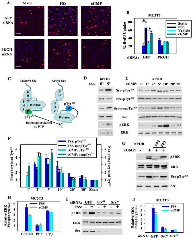
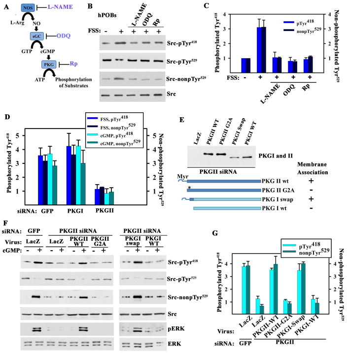
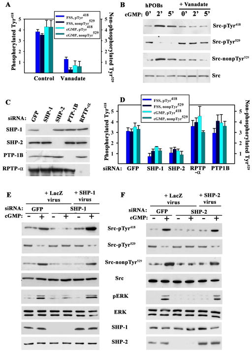
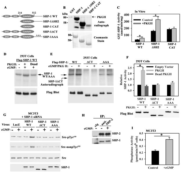
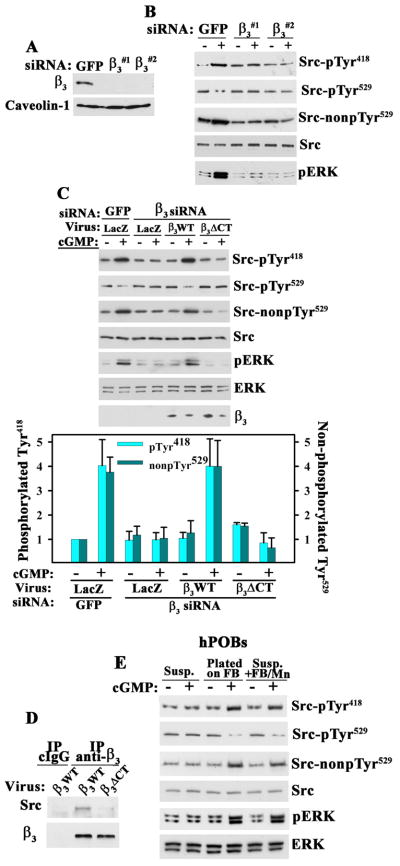
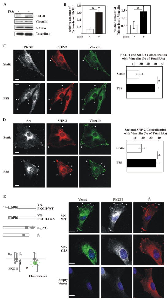
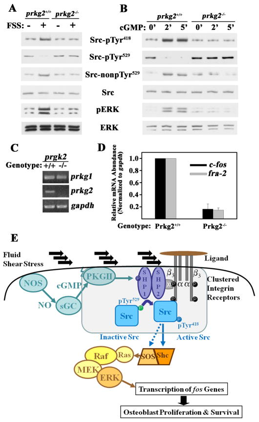
Mechanical stimulation is crucial for bone growth and remodeling, and fluid shear stress promotes anabolic responses in osteoblasts through multiple second messengers, including nitric oxide (NO). NO triggers production of cyclic guanosine 3',5'-monophosphate (cGMP), which in turn activates protein kinase G (PKG). We found that the NO-cGMP-PKG signaling pathway activates Src in mechanically stimulated osteoblasts to initiate a proliferative response. PKGII was necessary for Src activation, a process that also required the interaction of Src with β₃ integrins and dephosphorylation of Src by a complex containing the phosphatases SHP-1 (Src homology 2 domain-containing tyrosine phosphatase 1) and SHP-2. PKGII directly phosphorylated and stimulated SHP-1 activity, and fluid shear stress triggered the recruitment of PKGII, Src, SHP-1, and SHP-2 to a mechanosome containing β₃ integrins. PKGII-null mice showed defective Src and ERK (extracellular signal-regulated kinase) signaling in osteoblasts and decreased ERK-dependent gene expression in bone. Our findings reveal a convergence of NO-cGMP-PKG and integrin signaling and establish a previously unknown mechanism of Src activation. These results support the use of PKG-activating drugs to mimic the anabolic effects of mechanical stimulation of bone in the treatment of osteoporosis.


Conflict of interest statement

Competing interests: The authors declare no conflicts of interest.

Figures

Figure 1
Figure 1. Fluid Shear Stress- and cGMP-induced Osteoblast Proliferation and Erk Activation; Requirements for PKG II and Src
A. and B. MC3T3-E1 transformed murine osteoblast-like cells (MC3T3) were transfected with GFP or PKG II siRNAs, and kept static, exposed to fluid shear stress (FSS; 12 dynes/cm2), or treated with 100 μM 8-CPT-cGMP (cGMP) for 10 min. BrdU incorporation into DNA was detected by immunofluorescence, with >300 cells analyzed per condition. (B: mean of three experiments ± SEM; *p < 0.05). C. Schema depicting Src activation. D. and E. hPOBs were sham-treated, subjected to fluid shear stress, or treated with 100 μM cGMP for the indicated times. Western blots were analyzed with phospho-specific antibodies against Src-pTyr418, Src-pTyr529, or Erk-pTyr204, and antibodies recognizing Src with unphosphorylated Tyr529, total Src, or Erk (representative of two experiments). F. MC3T3 cells were sham-treated, subjected to fluid shear stress, or treated with 100 μM cGMP for the time indicated. Changes in Src phosphorylation were expressed relative to the amount of pTyr418 or pTyr529 found in sham-treated cells. (mean ± SEM; n=3; p < 0.05 for comparison between sham and 2 or 5 min time points). G. hPOBs were treated with 10 μM PP2 or PP3 for 1 hour and received 100 μM cGMP for 5 min (representative of two experiments). H. MC3T3 cells were pre-treated with PP2 or PP3 as in G, prior to exposure to either fluid shear stress or 100 μM cGMP for 5 min. Erk phosphorylation was expressed relative to sham-treated cells (mean ± SEM; n=3; *p < 0.05 for the comparison between PP2 versus control and PP2 versus PP3). I. and J. MC3T3 cells were transfected with siRNAs targeting GFP or two different sequences in Src, and were exposed to fluid shear stress or treated with 100 μM cGMP for 5 min. (J: mean ± SEM; n=3; *p < 0.05 for the comparison between siRNAs targeting Src versus GFP).
Figure 2
Figure 2. Src Activation by Membrane-bound PKG
A. Schema of the NO/cGMP/PKG signaling pathway with inhibitors of NOS, soluble guanylate cyclase (sGC), and PKG. B. hPOBs were sham-treated or exposed for 5 min to fluid shear stress (FSS; 12 dynes/cm2); some cells were pre-treated with 4 mM L-NAME, 10 μM ODQ, or 100 μM Rp-8-CPT-PET-cGMPS (Rp) for 1 hour. Src phosphorylation was determined as in Fig. 1D (representative of two experiments). C. MC3T3 cells were pre-treated as in B and exposed to fluid shear stress or 100 μM 8-CPT-cGMP (cGMP) for 5 min (mean ± SEM; n=3; * p < 0.05 compared to sham). D. MC3T3 cells were transfected with siRNAs specific for GFP, PKG I or PKG II, exposed to either fluid shear stress or 100 μM cGMP for 5 min, and analyzed as in Fig. 1F (mean ± SEM; n=3; p< 0.05 for the comparison between siRNA targeting PKG II versus GFP). E to G. MC3T3 cells were transfected with PKG II (or GFP) siRNA, and infected with adenoviral vectors encoding LacZ (control), siRNA-resistant wild-type (wt) or myristoylation-deficient PKG II (PKG II G2A), and wild-type or membrane-targeted PKG I (PKG I swap). The Western blot in E shows expression of PKG I and II constructs in PKG II siRNA-transfected MC3T3 cells (whole cell lysates); membrane association is indicated as determined by subcellular fractionation. In F, cells were treated with 100 μM cGMP for 5 min or left untreated and cells were analyzed as in Fig. 1E. G shows the mean ± SEM for three experiments (p < 0.05 for the comparison of PKG II wt or PKG I swap versus LacZ virus in PKG II siRNA-transfected cells).
Figure 3
Figure 3. Fluid Shear Stress- and cGMP-induced Src Activation Mediated by Shp-1/2
A. MC3T3 cells were treated with 10 μM vanadate for 1 hour prior to a 5 min exposure to fluid shear stress (FSS; 12 dynes/cm2) or 100 μM 8-CPT-cGMP (cGMP). Src phosphorylation was analyzed as in Fig. 1F (mean ± SEM; n=3; p < 0.05 for vanadate versus control). B. hPOBs were treated with vanadate and cGMP as in A (images are representative of three experiments) C. and D. MC3T3 cells were transfected with siRNAs targeting GFP, Shp-1, Shp-2, RPTP-α, or PTP-1B, and phosphatase expression was quantified by Western blotting (whole cell lysates for Shp-1, -2, and PTP-1B, and membrane lysates for RPTP-α). In D, cells were exposed to fluid shear stress or treated with 100 μM cGMP for 5 min and Src phosphorylation was analyzed as in Fig. 1F (mean ± SEM; n=3; p < 0.05 for the comparison between siRNAs targeting Shp-1 or Shp-2 versus GFP). E. and F. MC3T3 cells were transfected with siRNAs targeting GFP, Shp-1 (E) or Shp-2 (F). Cells were infected with adenovirus encoding LacZ, siRNA-resistant human Shp-1 (E), or Shp-2 (F), and received 100 μM cGMP for 5 min(representative of three experiments).
Figure 4
Figure 4. PKG II Phosphorylation of Shp-1/2 and Regulation of PTP Activity
A. Shp-1 constructs; AAA denotes alanine substitutions for Ser553, Ser556, and Ser557. B. PKG II phosphorylation of bacterially-expressed Shp-1 constructs in the presence of [γ-32PO4]ATP in vitro(representative of three experiments). C. Effect of PKG II phosphorylation on Shp-1 PTPase activity in vitro (mean ± SEM; n=3; * p < 0.05). D and E. 293T cells were co-transfected with Flag epitope-tagged Shp-1 constructs and empty vector, wild-type PKG II, or kinase-dead PKG II; cells were labeled with 32PO4 for 4 hours, and treated with 100 μM 8-CPT-cGMP (cGMP) for 10 min. Immunoprecipitates were analyzed by SDS-PAGE/autoradiography, and protein expression was determined by Western blotting (representative of three experiments). F. PTP activity of Flag-tagged Shp-1 constructs immunoprecipitated from 293T cells transfected and treated as in D and E. The activity of each construct was normalized to its activity in PKG-deficient cells (mean ± SEM; n=3; *p < 0.05). G. MC3T3 cells were transfected with Shp-1 siRNA and infected with adenovirus encoding LacZ or siRNA-resistant human Shp-1 wild-type, ΔCT, or AAA. Cells were exposed to 100 μM cGMP for 5 min and Src (de)phosphorylation was analyzed as in Fig. 1E (representative of two experiments). H. Coimmunoprecipitation of Shp-1 and -2 from MC3T3 cells treated with 100 μM cGMP for 5 min (representative of three experiments). I. PTP activity in anti-Shp-2 immunoprecipitates (containing Shp-1 and -2) from control MC3T3 cells and from cells treated with 100 μM cGMP for 5 min (mean ± SEM; n=3; *p < 0.05).
Figure 5
Figure 5. Integrin-dependence of Src Activation by cGMP/PKG II
A. and B. MC3T3 cells were transfected with siRNAs targeting GFP or two different sequences in integrin β3, and cell membranes were analyzed by Western blotting (A). Cells in B were exposed to 100 μM 8-CPT-cGMP (cGMP) for 5 min and analyzed as in Fig. 1E (representative of three experiments). C. MC3T3 cells transfected with siRNAs targeting GFP or β3 were infected with lentivirus encoding siRNA-resistant human β3WT or Src binding-deficient β3ΔCT. Cells were treated with 100 μM cGMP for 5 min and analyzed as in Fig. 1E. Expression of human β3 constructs was analyzed by Western blotting of whole cell lysates; the amount of endogenous mouse β3 is below detection. The bar graph shows the mean ± SEM of three experiments (p < 0.05 for β3WT versus LacZ virus in β3 siRNA-transfected, cGMP-treated cells). D. Coimmunoprecipitation of Src with wild-type human β3WT but not β3ΔCT from MC3T3 cells infected with β3-expressing lentivirus (representative of two experiments). Endogenous murine β3 is not efficient lt immunoprecipitated. E. hPOBs were kept in suspension (Susp.), allowed to attach to fibrinogen (FB)-coated dishes, or kept in suspension and stimulated with soluble fibrinogen (250 μg/ml) plus MnCl2 (2 mM) (Susp.+FB/Mn). After 1 hour, some cells received 100 μM cGMP for 5 min. Cells were analyzed as in Fig. 1E (representative of three experiments).
Figure 6
Figure 6. Characterization of a Src-containing Mechano-sensitive Complex in Osteoblasts
A. and B. Western blots of detergent-insoluble fractions isolated from static MC3T3 cells and from cells exposed for 5 min to orbital fluid shear stress (FSS; 120 rpm) (A). The bar graphs in B summarize three separate experiments (mean ± SEM; n=3; *p < 0.05 for static versus shear-stressed cells). C. and D. PKG II (white) and Shp-2 (red), or Src (white) and Shp-2 (red) colocalization with vinculin (green) was examined in static and shear-stressed (FSS) MC3T3 cells (C). Focal adhesions were defined as vinculin-positive membrane complexes > 0.5 μm size, and colocalization of PKG II and Shp-2, or Src and Shp-2 with focal adhesions was scored as a percentage of total focal adhesions (D shows the mean ± standard error of proportion for three experiments, with ~35 cells evaluated per condition; *p < 0.05 for static versus shear-stressed cells; bar 5 μm). D. Colocalization of αIIb3 integrins with PKG II monitored by bimolecular fluorescence complementation (BiFC) between αIIb-VC and wild-type PKG II-VN (VN-WT) or the membrane binding-deficient G2A mutant PKG II-VN (VN-G2A) in MC3T3 cells. All cells were transfected with human β3 and αIIb-VC (representative confocal images of three separate experiments; bar 5 μm).
Figure 7
Figure 7. Signaling Defect in PKG II-null Osteoblasts
A. Semi-quantitative RT-PCR for PKG I and II expression in primary calvarial osteoblasts isolated from one week-old PKG II−/− mice and their wild-type litter mates (representative of two experiments). B. and C. Wild-type and PKG II-null primary osteoblasts were stimulated with either fluid shear stress (FSS; 12 dynes/cm2) or 100 μM 8-CPT-cGMP (cGMP) for 5 min, and Src and Erk activation were analyzed as in Fig. 1E [representative of two (B) or three (C) experiments]. D. Tibial diaphyses were isolated from one week-old PKG II−/− mice and their wild-type litter mates, and c-fos, fra-2, and gapd mRNA amounts were measured by quantitative RT-PCR (mean ± SEM; n=3; * p < 0.05 for wild-type versus knockout mice). E. Model of Src and Erk activation by fluid shear stress via NO/cGMP/PKG II, depicting the assembly of a mechano-sensitive complex containing PKG II, Shp-1/2, and Src bound to the cytoplasmic tail of β3, as described in the text (x = docking protein). Activation of the Ras/Raf/MEK/Erk cascade by Src occurs via Shc-dependent and -independent pathways (33).

References

    1. Ozcivici E, Luu YK, Adler B, Qin YX, Rubin J, Judex S, Rubin CT. Mechanical signals as anabolic agents in bone. Nat Rev Rheumatol. 2010;6:50–59. - PMC - PubMed
    1. Bikle DD. Integrins, insulin like growth factors, and the skeletal response to load. Osteoporos Int. 2008;19:1237–1246. - PMC - PubMed
    1. Papachroni KK, Karatzas DN, Papavassiliou KA, Basdra EK, Papavassiliou AG. Mechanotransduction in osteoblast regulation and bone disease. Trends Mol Med. 2009;15:208–216. - PubMed
    1. McAllister TN, Frangos JA. Steady and transient fluid shear stress stimulate NO release in osteoblasts through distinct biochemical pathways. J Bone Miner Res. 1999;14:930–936. - PubMed
    1. Rangaswami H, Marathe N, Zhuang S, Chen Y, Yeh JC, Frangos JA, Boss GR, Pilz RB. Type II cGMP-dependent protein kinase mediates osteoblast mechanotransduction. J Biol Chem. 2009;284:14796–14808. - PMC - PubMed

Publication types

MeSH terms

Substances

LinkOut - more resources