. 2011 Apr;39(8):3093-102.
doi: 10.1093/nar/gkq1279. Epub 2010 Dec 21.

Nucleosome accessibility governed by the dimer/tetramer interface

Affiliations

Nucleosome accessibility governed by the dimer/tetramer interface

Vera Böhm et al. Nucleic Acids Res. 2011 Apr.

Abstract

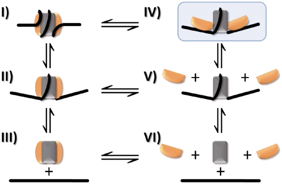
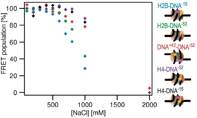
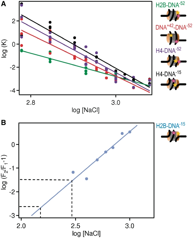
Nucleosomes are multi-component macromolecular assemblies which present a formidable obstacle to enzymatic activities that require access to the DNA, e.g. DNA and RNA polymerases. The mechanism and pathway(s) by which nucleosomes disassemble to allow DNA access are not well understood. Here we present evidence from single molecule FRET experiments for a previously uncharacterized intermediate structural state before H2A-H2B dimer release, which is characterized by an increased distance between H2B and the nucleosomal dyad. This suggests that the first step in nucleosome disassembly is the opening of the (H3-H4)(2) tetramer/(H2A-H2B) dimer interface, followed by H2A-H2B dimer release from the DNA and, lastly, (H3-H4)(2) tetramer removal. We estimate that the open intermediate state is populated at 0.2-3% under physiological conditions. This finding could have significant in vivo implications for factor-mediated histone removal and exchange, as well as for regulating DNA accessibility to the transcription and replication machinery.


Figures

Figure 1.
Figure 1.
Pathways for nucleosome (dis)assembly. A reaction mechanism depicting possible intermediate steps in the transition between nucleosome (I) and free DNA, H2A–H2B dimer and (H3–H4)2 tetramer (VI). DNA breathing (II), as first described by Widom and colleagues (38) increases the accessibility of DNA to protein binding. Disassembly has been proposed to occur either through a sequential mechanism (I→V→VI) or a one-step release of the histone octamer (I→III→VI). State IV (shaded in blue) represents a previously undetected open state in which the interactions between the H2A–H2B dimer and (H3–H4)2 tetramer are partially lost.
Figure 2.
Figure 2.
Fluorescently labeled nucleosomes. (A) Illustration of the extended 170 bp nucleosomal DNA showing the positions of the donor fluorophore alexa 488 (green circles) and acceptor fluorophores alexa 594 (red circles) relative to the nucleosome dyad axis. (B) Top and side views of the nucleosome crystal structure for visualization of fluorophore positions and FRET distances. H2A is shown in yellow, H2B in red, H3 in blue, H4 in green. The fluorophores are depicted with green and red circles corresponding to donor (alexa 488) and acceptor (alexa 594). Depending on perspective, some fluorophore positions are hidden and therefore not labeled in the crystal structure. In the top view, only fluorophores on one of the two H2B and on one of the two H4 are marked. In the side view, the fluorophore on position −15 on the DNA and the fluorophores on both H4 are not marked.
Figure 3.
Figure 3.
(H2A–H2B) dimers dissociate before (H3–H4)2 tetramer as shown by FCS. Diffusion coefficients of H2B-, H4- and DNA-labeled nucleosome samples (green, black and red, respectively) and labeled DNA (orange) relative to a standard (alexa 488) as a function of [NaCl] (error bars represent standard deviation from 10 measurements each). The sequential increase of the diffusion coefficients of H2B- and H4-labeled nucleosome samples indicates that H2A–H2B dissociates from the nucleosomal complex at lower [NaCl] than (H3–H4)2.
Figure 4.
Figure 4.
spFRET can be used to measure intranucleosomal conformational changes. (A) Example of a proximity ratio distribution for DNA+42–DNA−52, H2B–DNA−52, H2B–DNA−15, H4–DNA−52 and H4–DNA−15 at 150 mM NaCl. Cartoons of nucleosomes indicate the relative locations of labels on the nucleosome. The FRET population represents intact nucleosomes. The number of NoFRET bursts is low. The fit of FRET population is shown in black, fits for subpopulations of the FRET fraction (if present) are shown in different shades of blue. In intact nucleosomes, subpopulations arise if the distances between the fluorophore on the DNA and the fluorophores on the two copies of the labeled histones differ (sSupplementary Data 1.2). (B) Examples of proximity ratio distributions for each construct (as above) at 400 mM (green), 600 mM (blue), 800 mM (black) and 1200 mM (red) NaCl. Elevated [NaCl] causes an increase of the NoFRET population at the cost of the FRET population indicating that the distance between the fluorophores increased above the distance for energy transfer. Other than H4–DNA−15, all constructs displayed changes in the proximity ratio distribution of the FRET population upon increase of [NaCl] which are related to structural changes within the nucleosome.
Figure 5.
Figure 5.
spFRET reveals an intermediate open conformation before H2A–H2B dimer dissociation from the DNA. Plot showing fraction of the FRET population as a function of the [NaCl] for H2B–DNA−15 (blue), H2B–DNA−52 (green), DNA+42–DNA−52 (red), H4–DNA−52 (violet) and H4–DNA−15 (black). Each point represents an independent experiment. Errors were estimated based on the quality of the Gaussian fits of the proximity ratio distributions. Cartoons of nucleosomes indicating the relative locations of labels on the nucleosome, together with the c1/2 values derived from the sigmoidal curves are also shown, using the same color scheme. Donor labels are shown in yellow, acceptor labels are shown in magenta. From the sequence of the loss of FRET between the different nucleosome subunits, a model for disassembly can be derived (see text).
Figure 6.
Figure 6.
Nucleosome assembly follows the reverse pathway as disassembly. The fraction of FRET population as a function of [NaCl] measured during reconstitution of H2B–DNA−15 (blue), H2B–DNA−52 (green), DNA+42–DNA−52 (red), H4–DNA−52 (violet) and H4–DNA−15 (black). Cartoons of nucleosomes indicate the relative locations of labels on the nucleosome. The experimental error is estimated to be <6%. For clearness of the representation, error bars are not depicted. Upon lowering the [NaCl], FRET appears in the reverse order as it disappears during NaCl induced disassembly, indicating that the reconstitution follows the same mechanism as disassembly.
Figure 7.
Figure 7.
The number of ion pairs between histones and DNA can be derived from the salt dependence of the FRET fraction. (A) Equilibrium constants (K) for histone dissociation as a function of NaCl concentration for H2B–DNA−52 (green), DNA+42–DNA−52 (red), H4–DNA−52 (violet) and H4–DNA−15 (black). Cartoons of nucleosomes indicate the relative locations of labels on the nucleosome. K-values were calculated from the fraction of intact nucleosomes (Figure 5). The number of ions pairs between histones and DNA can be derived from curve fitting (as described in Supplementary Data 2.8, results see Supplementary Table S3). Approximately twice as many ion pairs are broken upon (H3–H4)2 than upon H2A–H2B dissociation. (B) Plot for the determination of the number of ion pairs involved in the opening of the H2A–H2B/(H3–H4)2 interface [see SupplementaryData, Equation (7)]. Data points were calculated from the fraction of intact nucleosomes of H2B–DNA−15 and H2B–DNA−52 (Figure 5). The transition requires the breaking of 4 ± 1 ion pairs as required from the slope of the fit. From extrapolation to physiological salt concentrations (150–300 mM NaCl, indicated by the dashed lines) the fraction of open nucleosomes occupied at physiological salt can be estimated to 0.2–3%.

References

    1. Luger K, Mäder AW, Richmond RK, Sargent DF, Richmond TJ. Crystal structure of the nucleosome core particle at 2.8 A resolution. Nature. 1997;389:251–260. - PubMed
    1. Van Holde KE. Chromatin. New York: Springer; 1988.
    1. Akey CW, Luger K. Histone chaperones and nucleosome assembly. Curr. Opin. Struct. Biol. 2003;13:6–14. - PubMed
    1. Lusser A, Kadonaga JT. Strategies for the reconstitution of chromatin. Nat. Methods. 2004;1:19–26. - PubMed
    1. Wilhelm FX, Wilhelm ML, Erard M, Duane MP. Reconstitution of chromatin: assembly of the nucleosome. Nucleic Acids Res. 1978;5:505–521. - PMC - PubMed

Publication types

MeSH terms