. 2011 Feb;25(2):253-68.
doi: 10.1210/me.2010-0318. Epub 2010 Dec 22.

CCAAT/enhancer-binding proteins (C/EBP)-α and -β are essential for ovulation, luteinization, and the expression of key target genes

Affiliations

CCAAT/enhancer-binding proteins (C/EBP)-α and -β are essential for ovulation, luteinization, and the expression of key target genes

Heng-Yu Fan et al. Mol Endocrinol. 2011 Feb.

Abstract

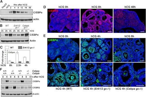
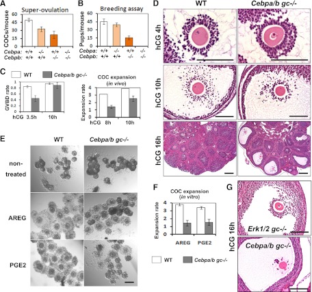
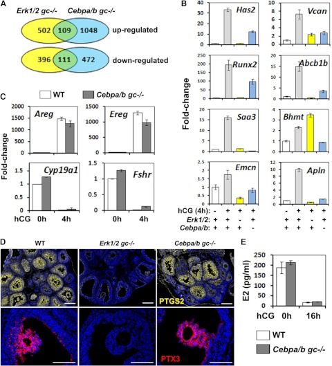
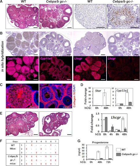
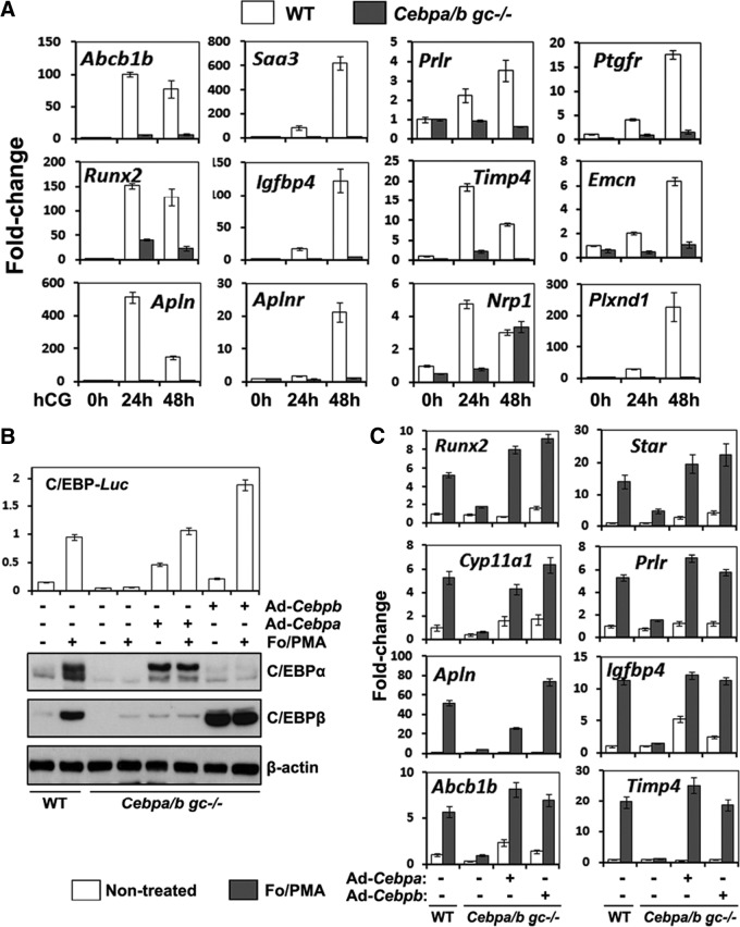
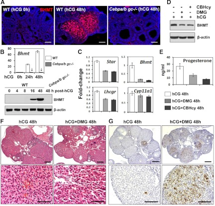
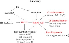
LH activation of the epidermal growth factor receptor/RAS/ERK1/2 pathway is essential for ovulation and luteinization because granulosa cell (GC) depletion of ERK1/2 (ERK1/2(gc)(-/-) mice) renders mice infertile. As mediators of ERK1/2-dependent GC differentiation, the CCAAT/enhancer-binding proteins, (C/EBP)α and C/EBPβ, were also disrupted. Female Cebpb(gc)(-/-) mutant mice, but not Cebpa(gc)(-/-) mice, were subfertile whereas Cebpa/b(gc)(-/-) double-mutant females were sterile. Follicles failed to ovulate, ovaries were devoid of corpora lutea, luteal cell marker genes (Lhcgr, Prlr, Ptgfr, Cyp11a1, and Star) were absent, and serum progesterone levels were low. Microarray analyses identified numerous C/EBPα/β target genes in equine chorionic gonadotropin (eCG)-human (h)CG-treated mice. At 4 h post-hCG, a subset (19%) of genes altered in the Cebpa/b-depleted cells was also altered in ERK1/2-depleted cells; hence they are common effectors of ERK1/2. Additional genes down-regulated in the Cebpa/b-depleted cells at 8 and 24 h post-hCG include known (Akr1b7, Runx2, Star, Saa3) and novel (Abcb1b, Apln, Igfbp4, Prlr, Ptgfr Timp4) C/EBP targets and effectors of luteal and vascular cell development. Bhmt, a gene controlling methionine metabolism and thought to be expressed exclusively in liver and kidney, was high in wild-type luteal cells but totally absent in Cebpa/b mutant cells. Because numerous genes potentially associated with vascular development were suppressed in the mutant cells, C/EBPα/β appear to dictate the luteinization process by also controlling genes that regulate the formation of the extensive vascular network required to sustain luteal cells. Thus, C/EBPα/β mediate the terminal differentiation of GCs during the complex process of luteinization.


Figures

Fig. 1.
Fig. 1.
Expression of C/EBPα and -β in preovulatory follicles. A, Western blots show the expression levels of C/EBPα protein in ovarian lysates of WT mice, before and after hCG treatment. B, Western blots show the expression levels of C/EBPα protein in ovarian lysates of WT, Erk½gc−/−, and Cebpa/bgc−/− mice, before and after hCG treatment. For each time point, ovarian lysates were made from three mice and pooled together. C, Real-time RT-PCR shows the expression levels of Cepba mRNA in granulosa cells collected from WT and Erk½gc−/− mice. D and E, Immunofluorescent staining shows the expression patterns of C/EBPβ (D) and C/EBPα (E) in ovaries of eCG-primed (44–48 h) 23-d-old immature mice before and after hCG treatment. (scale bar, 150 μm for all the images). F, Western blots show the expression levels of C/EBPα and -β protein in ovarian lysates of eCG-primed (44–48 h) WT, Cebpagc−/−, Cebpbgc−/−, and Cebpa/bgc−/− mice, before and after hCG treatment.
Fig. 2.
Fig. 2.
The Cebpa/bgc−/− mice cannot ovulate and are completely infertile. A and B, Fertility was examined by treating immature mice of each genotype with a superovulatory regimen of homones (A) and by breeding 6-wk-old females of indicated genotypes with fertile WT males for 6 months (n = 6 for each genotype) (B). Ovulated oocytes were collected from oviducts and counted at 16 h after hCG injections. (n = 8∼10 for each genotype.). C (left panel), The rate of oocyte GVBD was determined by counting the number of oocytes exhibiting GVBD in ovarian sections of WT and Cebpa/bgc−/− mice at 3.5 h and 10 h post-hCG (n = 6∼8 for each group); C (Right panel), The extent of COC expansion was determined as described in detail previously (74, 75) 0 = no expansion; 4 = maximum expansion. D, H&E staining shows the ovarian histology of 23-d-old WT and Cebpa/bgc−/− mice treated with eCG (48 h)+hCG (4 h, 10 h, and 16 h). Scale bar, 150 μm for all images. E, In vitro COC expansion assay. Fully grown COCs were isolated from WT and Cebpa/bgc−/− mice at 44–48 h after eCG treatment and cultured in COC medium (30 COCs/100 μl medium). Cumulus expansion was induced by an overnight treatment of AREG (100 ng/ml) or prostaglandin E2 (500 ng/ml). Scale bar, 150 μm. F, The extent of COC expansion in vitro was determined by quantification of expansion index. G, H&E staining shows the COC morphology of 23-d-old Erk½gc−/− and Cebpa/bgc−/− mice treated with eCG (48 h)+hCG (16 h). Scale bar, 70μm.
Fig. 3.
Fig. 3.
Microarray analyses identified LH-target genes regulated by C/EBPα and -β at the early stages of ovulation. A, Microarray analyses identified the genes up- and down-regulated in GCs isolated from Erk½gc−/− and Cebpa/bgc−/− mice at 4 h post-hCG. Approximately 19% of the genes were regulated in a similar pattern in both Erk½gc−/− and Cebpa/bgc−/− GCs. B and C, Real-time RT-PCR shows the mRNA expression levels of selected genes in GCs of WT, Erk½gc−/−, and Cebpa/bgc−/− mice treated with eCG (48 h)+hCG (4 h). D, Immunofluorescent staining shows the expression of PTGS2 (yellow) and PTX3 (red) in preovulatory follicles of WT, Erk½gc−/−, and Cebpa/bgc−/− mice treated with eCG (48 h)+hCG (4 h). Scale bar, 150μm. E, Serum estradiol levels in 25-d-old WT and Cebpa/bgc−/− mice before and after hCG treatment (0 h and 16 h) (n = 5 for each treatment.)
Fig. 4.
Fig. 4.
The Cebpa/bgc−/− mice exhibit severe defects in luteinization. A, Hematoxylin and eosin staining and TUNEL assay show the histology and apoptosis of luteal cells (48 h after hCG injection) in the ovaries of WT and Cebpa/bgc−/− mice. Scale bar, 150 μm. B, In situ hybridization shows the expression of mRNA encoding Cyp11a1 and Lhcgr in WT and Cebpa/bgc−/− ovaries at 48 h after hCG treatment. Histology of the ovaries is shown by hematoxylin staining (bright-field images); localization of Cyp11a1 and Lhcgr mRNAs is shown by dark-field images captured with Zeiss Axioplan microscope. Scale bar, 150μm. C, Immunostaining shows the presence of collgen IV, an endothelial cell marker, in the vascularized CL present in WT but not in Cebpa/bgc−/− mutant ovaries at 48 h after hCG. Scale bar, 150 μm. D, Real-time RT-PCR shows the expression of CL marker genes Star and Cyp11a1 in GCs and luteal cells of WT and Cebpa/bgc−/− mice before (0 h) and after (48 h) hCG treatment, respectively, and Lhcgr in cells isolated from WT and Cebpa/bgc−/− mice before (0 h) and after (8 h), and 48 h hCG treatment. (n = 3 for each group.) E, Hematoxylin and eosin staining shows the ovarian histology of 8-wk-old cycling WT and Cebpa/bgc−/− females. Scale bar, 150 μm. F, Days of estrus indicated by vaginal cytology in cycling WT and Cebpa/bgc−/− females (3 months old). G, Serum progesterone levels in 25-d-old WT and Cebpa/bgc−/− mice before and after hCG treatment (0 h, 48 h, and 72 h) (n = 5 for each treatment).
Fig. 5.
Fig. 5.
Microarray analyses identify novel C/EBP target genes that regulate ovulation and luteinization. A, Granulosa/luteal cells were isolated from WT and Cebpa/bgc−/− mice before and after hCG treatment (0 h, 24 h, and 48 h) (n = 3 for each treatment). Total RNA was isolated, and the expression levels of indicated genes were determined by real-time RT-PCR. B, GCs were isolated from eCG primed (24 h) 23-d-old immature mice (WT and Cebpa/bgc−/−) and cultured for 24 h. At that time, Cebpa/bgc−/− GCs were washed and infected with adenoviral vectors encoding either C/EBPα or C/EBPβ. After 4 h, the cells were treated with For (10 μm) plus PMA (20 nm) for another 20 h. Western blots show the expression levels of C/EBPα and C/EBPβ proteins. C, GCs were cultured, and infected with adenoviral vectors as in panel B. Then the cells were treated with For/PMA for another 24 h. Expression of indicated C/EBP-target genes was detected by real-time RT-PCR.
Fig. 6.
Fig. 6.
Bhmt is a novel C/EBP-regulated gene that is required for the maintenance of luteal cell functions. A and B, Immunofluorescent staining (A) and real-time RT-PCR and Western blot (B) analyses show the expression of BHMT protein and mRNA in ovaries of WT and Cebpa/bgc−/− mice, before and after hCG treatment (48 h). Arrows in panel B indicate the extremely low expression levels of Bhmt mRNA in the ovaries of the Cebpa/bgc−/− mice. Scale bar, 150 μm. C, Quantitative RT-PCR shows the effects of BHMT-specific inhibitors, DMG or CBHcy, on the expression of selected luteal cell marker genes at 48 h post-hCG. D, Western blot shows the effect of DMG and CBHcy on the levels of BHMT protein in ovaries at 48 h post-hCG. E, Serum progesterone levels (48 h after hCG injection) of WT mice with or without treatment of BHMT inhibitors (n = 5 for each treatment). F and G, H&E staining (F) and TUNEL assay (G) show the histology and apoptosis of luteal cells (48 h after hCG injection) in WT mice treated with or without DMG treatment.
Fig. 7.
Fig. 7.
A schematic of C/EBP α/β functions in GCs. LH induces ovulation and luteinization by activating the cAMP/PKA pathway leading to expression of the EGF-like factors, AREG, epiregulin, and betacellulin. These factors then bind the EGFR to activate the RAS/ERK½ signaling cascade (8, 76). The results presented herein show that C/EBPα and -β are essential for LH induction of ovulation and luteinzation because they mediate specific events downstream of LH, PKA, and ERK½ (8, 18, 20, 77). By generating the Cebpa/bgc−/− double-mutant mice, analyzing gene profiling datasets and verifying target genes by in vitro approaches, we have identified specific genes that are potential targets (direct or indirect) of C/EBPα/β. These genes are involved in the formation and maintenance of CL, steroidogensis, and the vascularization of ruptured follicles.

References

    1. Fan HY, Liu Z, Cahill N, Richards JS. 2008. Targeted disruption of Pten in ovarian granulosa cells enhances ovulation and extends the life span of luteal cells. Mol Endocrinol 22:2128–2140 - PMC - PubMed
    1. Richards JS, Jonassen JA, Rolfes AI, Kersey K, Reichert LE., Jr 1979. Cyclic AMP, luteinizing hormone receptor, and progesterone during granulosa cell differentiation: effects of estradiol and follicle stimulating hormone. Endocrinology 104:765–773 - PubMed
    1. Richards JS, Midgley AR., Jr 1976. Protein hormone action: a key to understanding ovarian follicular and luteal cell development. Biol Reprod 14:82–94 - PubMed
    1. Salvador LM, Maizels E, Hales DB, Miyamoto E, Yamamoto H, Hunzicker-Dunn M. 2002. Acute signaling by the LH receptor is independent of protein kinase C activation. Endocrinology 143:2986–2994 - PubMed
    1. Sharma SC, Richards JS. 2000. Regulation of AP1 (Jun/Fos) factor expression and activation in ovarian granulosa cells: relation of JunD and Fra2 to terminal differentiation. J Biol Chem 275:33718–33728 - PubMed

Publication types

MeSH terms

Substances