. 2011 Mar;85(5):2024-36.
doi: 10.1128/JVI.01895-10. Epub 2010 Dec 22.

A single mutation responsible for temperature-sensitive entry and assembly defects in the VP1-2 protein of herpes simplex virus

Affiliations

A single mutation responsible for temperature-sensitive entry and assembly defects in the VP1-2 protein of herpes simplex virus

F Abaitua et al. J Virol. 2011 Mar.

Abstract

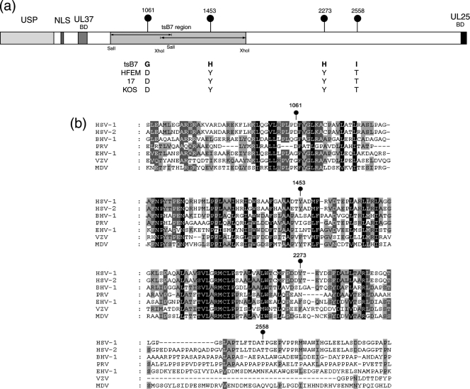
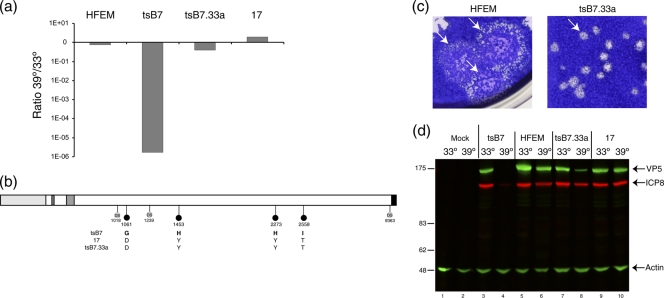
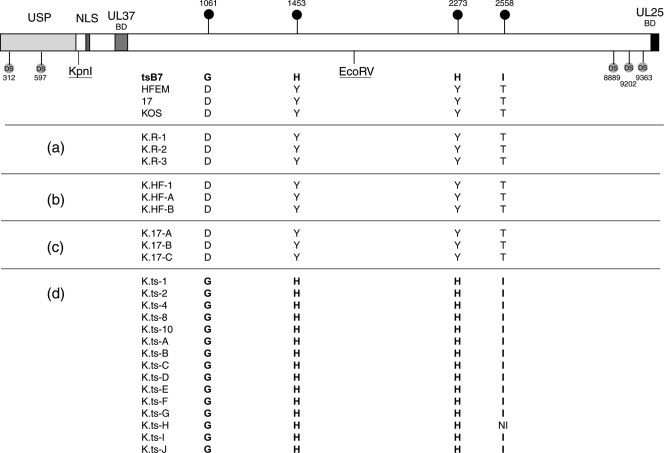
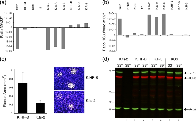
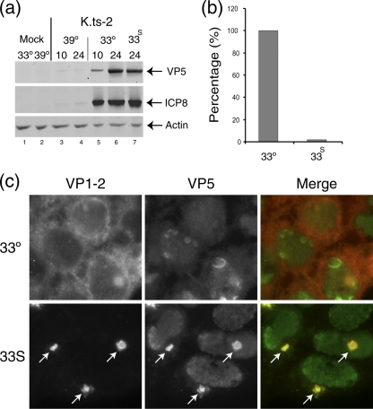
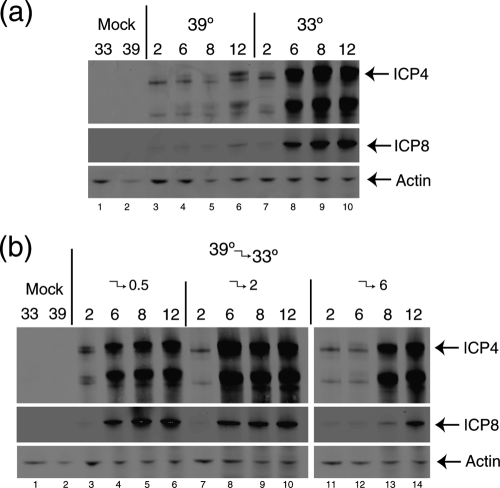
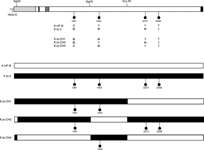
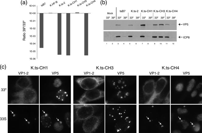
Evidence for an essential role of the herpes simplex virus type 1 (HSV-1) tegument protein VP1-2 originated from the analysis of the temperature-sensitive (ts) mutant tsB7. At the nonpermissive temperature (NPT), tsB7 capsids accumulate at the nuclear pore, with defective genome release and substantially reduced virus gene expression. We compared the UL36 gene of tsB7 with that of the parental strain HFEM or strain 17 and identified four amino acid substitutions, 1061D → G, 1453Y → H, 2273Y → H, and 2558T → I. We transferred the UL36 gene from tsB7, HFEM, or strain 17 into a KOS background. While KOS recombinants containing the HFEM or strain 17 UL36 gene exhibited no ts defect, recombinants containing the tsB7 UL36 VP1-2 exhibited a 5-log deficiency at the NPT. Incubation at the NPT resulted in little or no virus gene expression, though limited expression could be detected in a highly delayed fashion. Using shift-down regimes, gene expression recovered and recapitulated the time course normally observed, indicating that the initial block was in a reversible pathway. Using temperature shift-up regimes, a second defect later in the replication cycle was also observed in the KOS.ts viruses. We constructed a further series of recombinants which contained subsets of the four substitutions. A virus containing the wild-type (wt) residue at position 1453 and with the other three residues being from tsB7 VP1-2 exhibited wt plaquing efficiency. Conversely, a virus containing the three wt residues but the single Y → H change at position 1453 from tsB7 exhibited a 4- to 5-log drop in plaquing efficiency and was defective at both early and late stages of infection.


Figures

FIG. 1.
FIG. 1.
The tsB7 UL36 gene contains four amino acid substitutions. (a) The UL36 genes from tsB7, parental strain HFEM, and wt strain 17 were sequenced. Positions of amino acid substitutions (numbered with reference to the HFEM sequence [see Materials and Methods]) are indicated together with the residues at these positions in tsB7 or the other strains. Features of VP1-2 illustrated in the diagram include the ubiquitin-specific protease (USP), nuclear localization signal (NLS), UL37-binding domain (UL37BD), and conserved C-terminal UL25-binding domain (UL25BD). The positions of fragments used for marker rescue of tsB7 encompassed by SalI or XhoI sites are also indicated (3). (b) Sequence homology around the tsB7 substitutions in the VP1-2 proteins of alphaherpesvirus subfamily members. Alignments were made using ClustalX alignment software and illustrated using Genedoc. Residues with complete identity allowing for close chemical similarity are indicated in white text with a black background, over 85% by white text on dark gray, and over 50% by black text on light gray.
FIG. 2.
FIG. 2.
Construction of a recombinant tsB7 virus containing the wt UL36 gene. tsB7 DNA was cotransfected with a plasmid carrying the wt UL36 gene from strain 17, and the isolate tsB733a was further characterized. (a) Restoration of plaquing efficiency at 39°C. Plaque assay was performed on Vero cells infected at 33°C or 39°C with the HFEM, tsB7, tsB733a, and 17 viruses. The graph shows the relative ratio of the plaque formation of each virus. (b) The UL36 gene from tsB733a was subcloned and sequenced. Annotation is as for Fig. 1. DS, diagnostic nucleotide polymorphisms allowing sequence attribution. (c) Vero cells were infected with dilutions of HFEM and tsB733a viruses and incubated at 39°C, and plates were stained with crystal violet. Representative images of relative plaque size are shown. (d) Expression of candidate viral protein ICP8 and VP5 expression levels at 33°C and 39°C in Vero cells infected with tsB7, HFEM, tsB733a, and strain 17 at an MOI of 5. For quantitation using the LiCor Odyssey system, relative levels of each protein were normalized with actin levels.
FIG. 3.
FIG. 3.
UL36 sequence analysis of the KOS recombinant viruses. COS cells were cotransfected at 33°C with KΔUL36 DNA either alone or with pUC19 plasmids carrying UL36 genes from strain HFEM, 17, or tsB7. (KΔUL36 is deleted between the indicated KpnI and EcoRV sites.) Several isolates from each transfection were purified by successive single-plaque isolations on Vero cells at 33°C. The resulting viruses were amplified and purified viral DNA sequenced. The table shows for each isolate virus the identity of the residue at the position of the four substitutions in tsB7, organized per transfection group: (a) KΔUL36 transfection alone (i.e., revertant KOS viruses K.R-x); (b) KΔUL36 plus HFEM UL36 (K.HF-x); (c) KΔUL36 plus strain 17 UL36 (K.17-x); and (d) KΔUL36 plus tsB7 UL36 (K.ts-x). Each individual virus was assigned a specific number or letter. The positions of diagnostic polymorphic nucleotide sequences (DS) are also indicated, which aided in demonstrating the extent of recombined sequences. NI, no sequence information available.
FIG. 4.
FIG. 4.
Temperature sensitivity of plaque formation by K.ts recombinant viruses. (a) Several isolates of each group of KOS recombinant viruses were analyzed for plaque formation in Vero cells at 39°C versus 33°C. Each of the K.ts isolates exhibited a reduction of approximately 5 logs in plaque formation. (b) Complementation of plaque formation in HS30 cells. Plaque formation in Vero or HS30 cells was quantitated at 39°C and the ratio determined. tsB7 plaque formation was of the order of 3 log units higher in HS30 cells, a result also obtained with each of the K.ts isolates. Comparatively minor differences between Vero and HS30 cells were observed for the other strains. (c) Plaque size for K.HF-B and K.ts-2 viruses at 33°C. The K.ts-2 plaque size was approximately one-third of the area of the corresponding isolates containing the HFEM UL36 gene. Plaque size was estimated using Image Pro Plus software from approximately 50 plaques for each virus, with representative images shown by crystal violet staining. Error bars indicate standard deviations. (d) Temperature sensitivity of gene expression. Vero cells were infected (MOI, 5) with K.ts-2, K.HF-B, K.R-3, or wt KOS at 33°C or 39°C, and extracts collected at 24 h postinfection and analyzed for the accumulation of viral proteins VP5 and ICP8. Actin levels were measured as a loading control.
FIG. 5.
FIG. 5.
Postentry defect of K.ts-2. Monolayers of Hep2 cells were infected (MOI, 5) with K.ts-2 at 33°C, and at 10 h one set of cultures was maintained at 33°C while a duplicate set was shifted up to 39°C (33S). Incubation was continued and cultures harvested at 24 h postinfection. (a) Examination of candidate virus protein expression (ICP8 and VP5) expressed after continuous incubation at 33°C (lanes 5 and 6) or 39°C (lanes 3 and 4) or under the shift regime (33S) (lane 7). Actin levels were measured as loading control. (b) Parallel examination of total virus yield determined by titration on Vero cell monolayers at 33°C. The yield under the shift regime was reduced almost 2 log units compared to that at 33°C (set as 100%). (c) Altered localization of ts VP1-2 after temperature shift. Hep2 cells were infected with K.ts-2 at 33°C, and at 10 h postinfection were either maintained at 33°C or shifted up to 39°C. The cells were fixed at 16 h postinfection and the localization of VP1-2 and VP5 analyzed. At 39°C, pronounced relocalization of VP1-2, colocalizing with VP5 in a single juxtanuclear aggregate, is evident (arrows).
FIG. 6.
FIG. 6.
Reversible early defect in K.ts-2 upon temperature shift-down assay. Vero cells were infected with K.ts-2 (MOI, 5) continuously at 39°C or 33°C (a) or infected at 39°C and shifted down to 33°C at 0.5 h postinfection (lanes 3 to 6), 2 h postinfection (lanes 7 to 10) or 6 h postinfection (lanes 11 to 14) (b) and then harvested at the times indicated. Total times are indicated. Thus, e.g., for lanes 11 to 13, samples were at 39°C for 2 h, at 39°C for 6 h (lane 12), and at 39°C for 6 h, followed by 2 h at 33°C (lane13). Levels of representative immediate-early proteins (ICP4) and delayed-early proteins (ICP8) are shown together with actin levels as loading controls.
FIG. 7.
FIG. 7.
Construction of the KOS recombinant viruses with selected substitutions in VP1-2. VP1-2 genes were constructed by swapping segments of parental HFEM and tsB7 genes using the indicated restriction sites (see Materials and Methods), yielding genes with various combinations of substitutions (lollipops indicate the presence of a residue from tsB7 UL36, and the absence of a lollipop indicates wt residues at those positions). Recombinant viruses were then constructed as before by cotransfecting COS cells with KΔUL36 DNA with the various plasmids. Isolates from each cotransfection were purified by successive single-plaque isolations on Vero cells at 33°C. The resulting viruses were amplified and purified viral DNA sequenced at the relevant positions of the four substitutions. Viruses were named for the provenance of the UL36 plasmids from which they were constructed, i.e., K.ts-CHx.
FIG. 8.
FIG. 8.
Identification of a single residue change responsible for the ts phenotype of tsB7 and K.ts-2. (a) tsB7, K.ts-2 (containing the four changes present in tsB7 UL36), and variants with subsets of these changes were analyzed for plaque formation in Vero cells at 39°C versus 33°C. K.ts-CH1 and K.ts-CH4 exhibited a reduction of approximately 5 log units in plaque formation, while K.ts-CH3 (containing three of the four substitutions but wt at position 1453) exhibited no defect in plaque formation. (b) Temperature sensitivity of gene expression. Vero cells were infected (MOI, 5) with viruses as indicated at 33°C or 39°C, and extracts were collected at 24 h postinfection and analyzed for the accumulation of viral proteins VP5 and ICP8. Actin levels were measured as a loading control. (c) Altered localization of ts VP1-2 after temperature shift. Hep2 cells were infected with the indicated viruses at 33°C and at 10 h postinfection either maintained at 33°C or shifted up to 39°C. Cells were fixed at 16 h postinfection and the localization of VP1-2 and VP5 analyzed. At 39°C, pronounced relocalization of VP1-2, colocalizing with VP5 in a single juxtanuclear aggregate, is evident (arrows) for K.ts-CH1 and for K.ts-CH4, which contain, respectively, 1061D → G plus 1453Y → H or just 1453Y → H. No alteration in VP1-2 localization was observed for K.ts-CH3, which contains three tsB7 substitutions, but the wt residue at position 1453.

References

    1. Abaitua, F., and P. O'Hare. 2008. Identification of a highly conserved, functional nuclear localization signal within the N-terminal region of herpes simplex virus type 1 VP1-2 tegument protein. J. Virol. 82:5234-5244. - PMC - PubMed
    1. Abaitua, F., R. N. Souto, H. Browne, T. Daikoku, and P. O'Hare. 2009. Characterization of the herpes simplex virus (HSV)-1 tegument protein VP1-2 during infection with the HSV temperature-sensitive mutant tsB7. J. Gen. Virol. 90:2353-2363. - PubMed
    1. Batterson, W., D. Furlong, and B. Roizman. 1983. Molecular genetics of herpes simplex virus. VIII. Further characterization of a temperature-sensitive mutant defective in release of viral DNA and in other stages of the viral reproductive cycle. J. Virol. 45:397-407. - PMC - PubMed
    1. Batterson, W., and B. Roizman. 1983. Characterization of the herpes simplex virion-associated factor responsible for the induction of alpha genes. J. Virol. 46:371-377. - PMC - PubMed
    1. Benyesh-Melnick, M., P. A. Schaffer, R. J. Courtney, J. Esparza, and S. Kimura. 1975. Viral gene functions expressed and detected by temperature-sensitive mutants of herpes simplex virus. Cold Spring Harbor Symp. Quant. Biol. 39:731-746. - PubMed

Publication types

Substances