. 2011 Mar;85(5):2325-32.
doi: 10.1128/JVI.01791-10. Epub 2010 Dec 22.

The herpes simplex virus type 1 latency-associated transcript can protect neuron-derived C1300 and Neuro2A cells from granzyme B-induced apoptosis and CD8 T-cell killing

Affiliations

The herpes simplex virus type 1 latency-associated transcript can protect neuron-derived C1300 and Neuro2A cells from granzyme B-induced apoptosis and CD8 T-cell killing

Xianzhi Jiang et al. J Virol. 2011 Mar.

Abstract

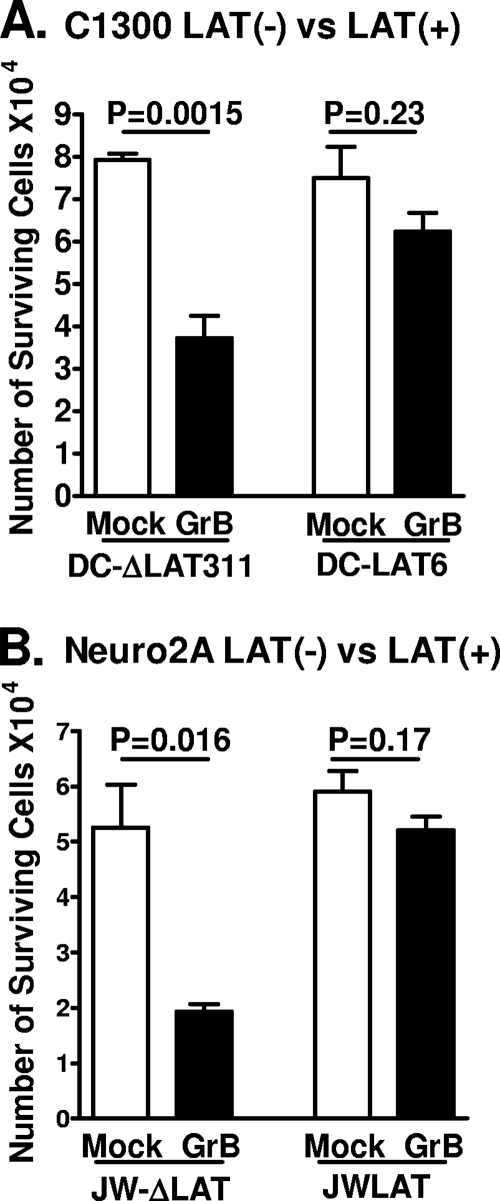
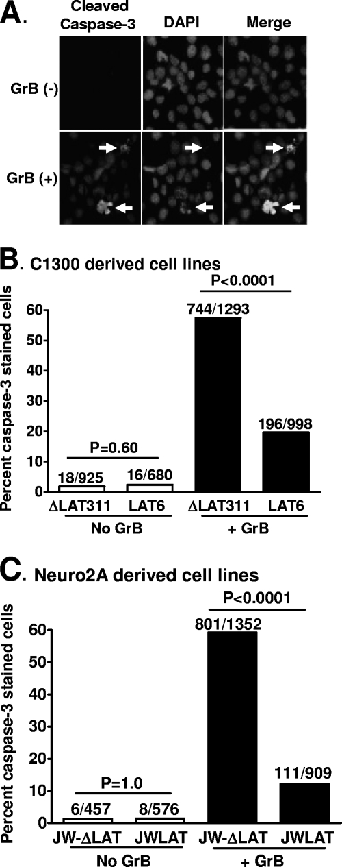
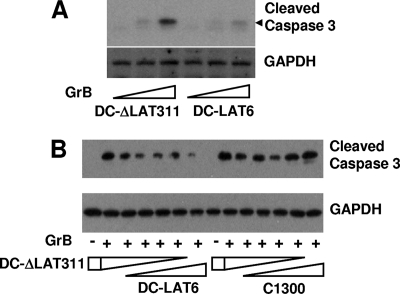
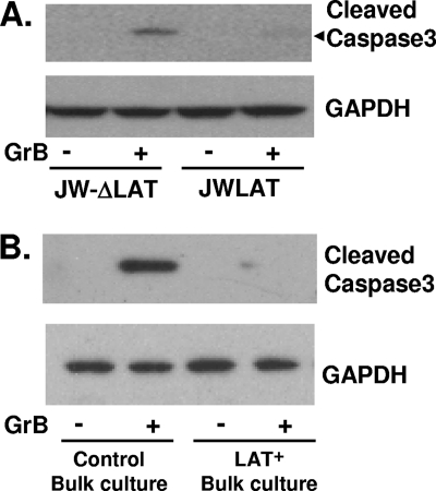
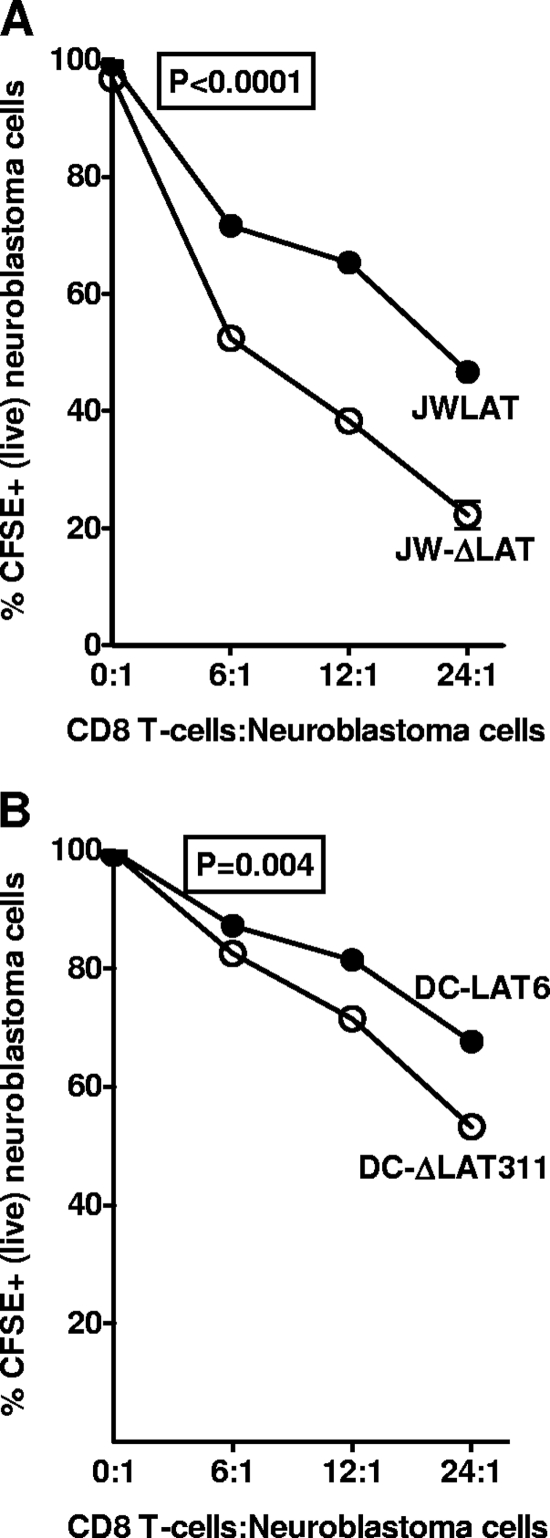
The herpes simplex virus type 1 (HSV-1) latency-associated transcript (LAT) is the only HSV-1 gene transcript abundantly expressed throughout latency. LAT null mutants have a significantly reduced reactivation phenotype. LAT's antiapoptosis activity is the major LAT factor involved in supporting the wild-type reactivation phenotype. During HSV-1 latency, some ganglionic neurons are surrounded by CD8 T cells, and it has been proposed that these CD8 T cells help maintain HSV-1 latency by suppressing viral reactivations. Surprisingly, despite injection of cytotoxic lytic granules by these CD8 T cells into latently infected neurons, neither apoptosis nor neuronal cell death appears to occur. We hypothesized that protection of latently infected neurons against cytotoxic CD8 T-cell killing is due to LAT's antiapoptosis activity. Since CD8 T-cell cytotoxic lytic granule-mediated apoptosis is critically dependent on granzyme B (GrB), we examined LAT's ability to block GrB-induced apoptosis. We report here that (i) LAT can interfere with GrB-induced apoptosis in cell cultures, (ii) LAT can block GrB-induced cleavage (activation) of caspase-3 both in cell culture and in a cell-free in vitro cell extract assay, and (iii) LAT can protect C1300 and Neuro2A cells from cytotoxic CD8 T-cell killing in vitro. These findings support the hypothesis that LAT's antiapoptosis activity can protect latently infected neurons from being killed by CD8 T-cell lytic granules in vivo.


Figures

FIG. 1.
FIG. 1.
Schematic representation of the LAT regions expressed in C1300- and Neuro2A-derived stable cell lines. (A) The LAT promoter (hollow rectangle) containing the TATA box, the start of LAT transcription (nt 1), the beginning (nt 661) and end (nt 2617) of the stable 2-kb LAT intron (solid rectangle), and the end of the primary LAT sequence (nt 8324) are shown. (B) The DC-LAT6 insert contains the first 3,225 nt of the primary LAT driven by the LAT promoter, cloned into the NotI site of plasmid pSL301. (C) The DC-ΔLAT311 insert is identical to the DC-LAT6 insert except for a PstI-PstI deletion that deletes key LAT promoter elements, including the TATA box, resulting in only trace transcription of LAT (presumably from a cellular promoter). (D) The JWLAT insert contains the first 1,499 nt of LAT driven by the LAT promoter, cloned into the BamHI site of plasmid pPUR. (E) The JW-ΔLAT insert is the blank plasmid used for JWLAT cells. DC-LAT6 and DC-ΔLAT311 inserts are in C1300 cells and were described previously (9). JWLAT and JW-ΔLAT inserts are in Neuro2A cells and were constructed as described in Materials and Methods. The Neuro2A-based JWLAT and JW-ΔLAT cell lines and the C1300-based DC-LAT6 and DC-ΔLAT311 cell lines were generated at different times and by different people.
FIG. 2.
FIG. 2.
LAT expression in DC-LAT6 and JWLAT cells decreased GrB-induced cell death. (A) Subconfluent monolayers of LAT+ DC-LAT6 and LAT DC-ΔLAT311 cells in 24-well plates were treated with 6 μg/ml of mouse GrB in serum-free medium for 24 h. The number of viable cells in each individual well was determined for at least 3 wells/group by trypan blue exclusion. The average number of viable cells per well ± the standard deviation (SD) for each group is shown. (B) Survival rates for LAT+ JWLAT cells and LAT JW-ΔLAT cells were determined as described in the legend to panel A, except that GrB treatment in serum-free medium was done for 6 h rather than 24 h, following which the cells were fed with medium containing 10% serum and incubated overnight. Panel B shows results for 1 of 3 independently performed experiments, all with similar outcomes. Taken together, panels A and B therefore represent the results of 4 independent experiments, each done in triplicate.
FIG. 3.
FIG. 3.
LAT expression in DC-LAT6 and JWLAT cells decreased GrB-induced apoptosis as judged by intracellular staining for cleaved (activated) caspase-3. Subconfluent cell monolayers on coverslips were treated with GrB as described in the legend to Fig. 2 [GrB (+)] or mock treated [GrB (−)] and then stained for cleaved caspase-3 by using a rabbit antibody specific for activated, cleaved caspase-3. (A) Examples of activated-caspase-3 staining in LAT LATΔ311 cells with and without GrB treatment are shown. Arrows indicate some cells with obvious apoptotic morphologies. (B) The percentages of DC-ΔLAT311 and DC-LAT6 apoptotic cells as judged by positivity for cleaved-caspase-3 staining are shown (on the y axis). The numbers above each bar are the number of cleaved-caspase-3-positive cells and the total number of cells counted. P values were determined by a two-sided chi-square test. (C) The percentages of cleaved-caspase-3-positive JWLAT and JW-ΔLAT cells are shown. The results shown for JWLAT and JW-ΔLAT cells are representative of results from 2 independent experiments.
FIG. 4.
FIG. 4.
GrB-induced caspase-3 cleavage in extracts of DC-LAT6 and DC-ΔLAT311 cells. (A) Cell extracts prepared as described in Materials and Methods were treated with increasing amounts of GrB in vitro as indicated (bottom) and analyzed by Western blotting using cleaved-caspase-3-specific rabbit antibody. Doses of GrB were 0, 0.025, and 0.05 μg/ml for 2 h. The predicted location of the cleaved-caspase-3 band is indicated by the black arrowhead. GAPDH is a loading control. The results shown are representative of results from 3 independent experiments. (B) Extracts from DC-ΔLAT311 and DC-LAT6 cells (left) or extracts from DC-ΔLAT311 and parental C1300 cells (right) were mixed together at various ratios (1:0, 4:1, 3:2, 2:3, 1:4, and 0:1) and treated with 0.2 μg/ml of GrB (+) or left untreated (−).
FIG. 5.
FIG. 5.
GrB-induced caspase-3 cleavage in extracts of JWLAT and JW-ΔLAT cells. (A) Extracts of JWLAT and JW-ΔLAT cells were prepared, treated with 0.05 μg/ml of GrB, and analyzed by Western blotting as described in the legend to Fig. 4A. The results shown are representative of results from 2 independent experiments. (B) Extracts of bulk Neuro2A cell cultures transfected with either the JWLAT or the JW-ΔLAT plasmid (see the text) were prepared and treated as described in the legend to panel A.
FIG. 6.
FIG. 6.
LAT inhibits cell death caused by mouse CD8 T cells. CD8 T cells from mice immunized with Neuro2A cells were stimulated in vitro, and increasing amounts were incubated with LAT+ or LAT cells that had been prelabeled with CFSE as described in Materials and Methods. P values were determined by a two-sided paired Student t test.

References

    1. Adrain, C., B. M. Murphy, and S. J. Martin. 2005. Molecular ordering of the caspase activation cascade initiated by the cytotoxic T lymphocyte/natural killer (CTL/NK) protease granzyme B. J. Biol. Chem. 280:4663-4673. - PubMed
    1. Ahmed, M., M. Lock, C. G. Miller, and N. W. Fraser. 2002. Regions of the herpes simplex virus type 1 latency-associated transcript that protect cells from apoptosis in vitro and protect neuronal cells in vivo. J. Virol. 76:717-729. - PMC - PubMed
    1. Aubert, M., and J. A. Blaho. 2001. Modulation of apoptosis during herpes simplex virus infection in human cells. Microbes Infect. 3:859-866. - PubMed
    1. Best, S. M. 2008. Viral subversion of apoptotic enzymes: escape from death row. Annu. Rev. Microbiol. 62:171-192. - PMC - PubMed
    1. Branco, F. J., and N. W. Fraser. 2005. Herpes simplex virus type 1 latency-associated transcript expression protects trigeminal ganglion neurons from apoptosis. J. Virol. 79:9019-9025. - PMC - PubMed

Publication types

MeSH terms

Substances