. 2011 Feb 25;286(8):6423-32.
doi: 10.1074/jbc.M110.180661. Epub 2010 Dec 22.

Protein kinase A-alpha directly phosphorylates FoxO1 in vascular endothelial cells to regulate expression of vascular cellular adhesion molecule-1 mRNA

Affiliations

Protein kinase A-alpha directly phosphorylates FoxO1 in vascular endothelial cells to regulate expression of vascular cellular adhesion molecule-1 mRNA

Ji-Won Lee et al. J Biol Chem. .

Abstract

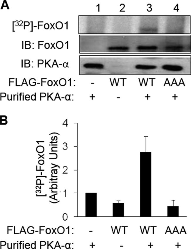
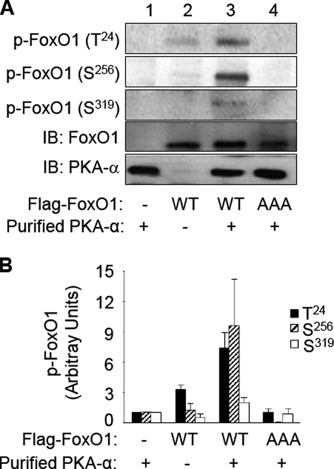
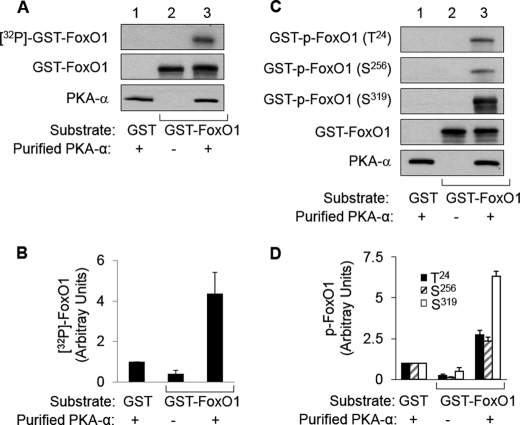
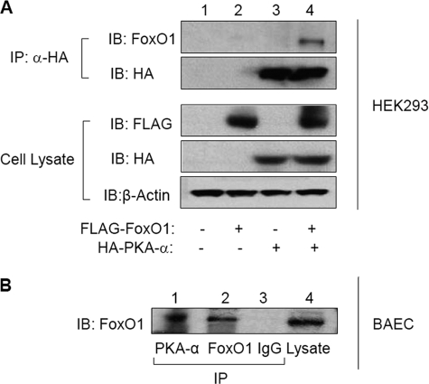
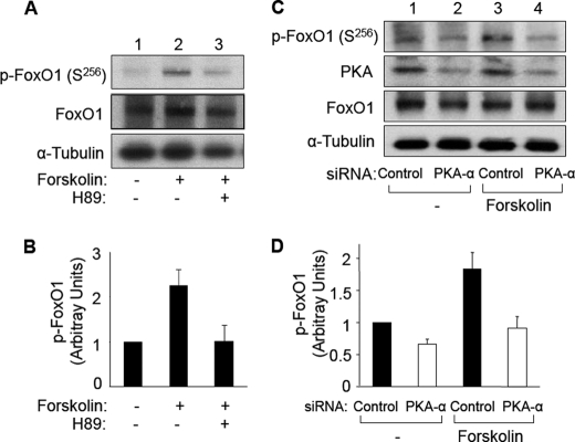
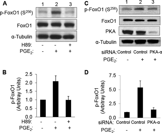
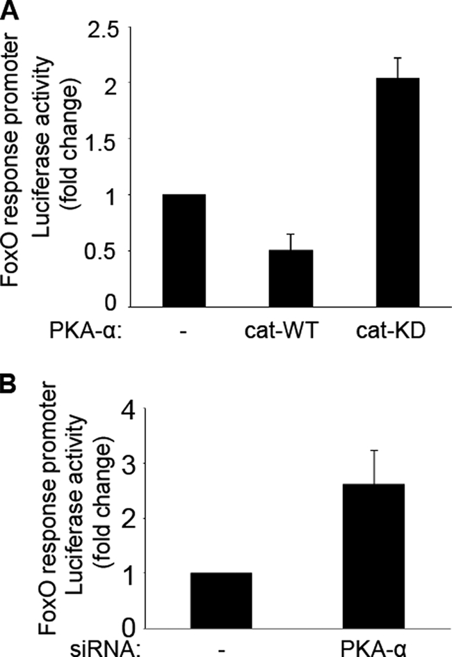
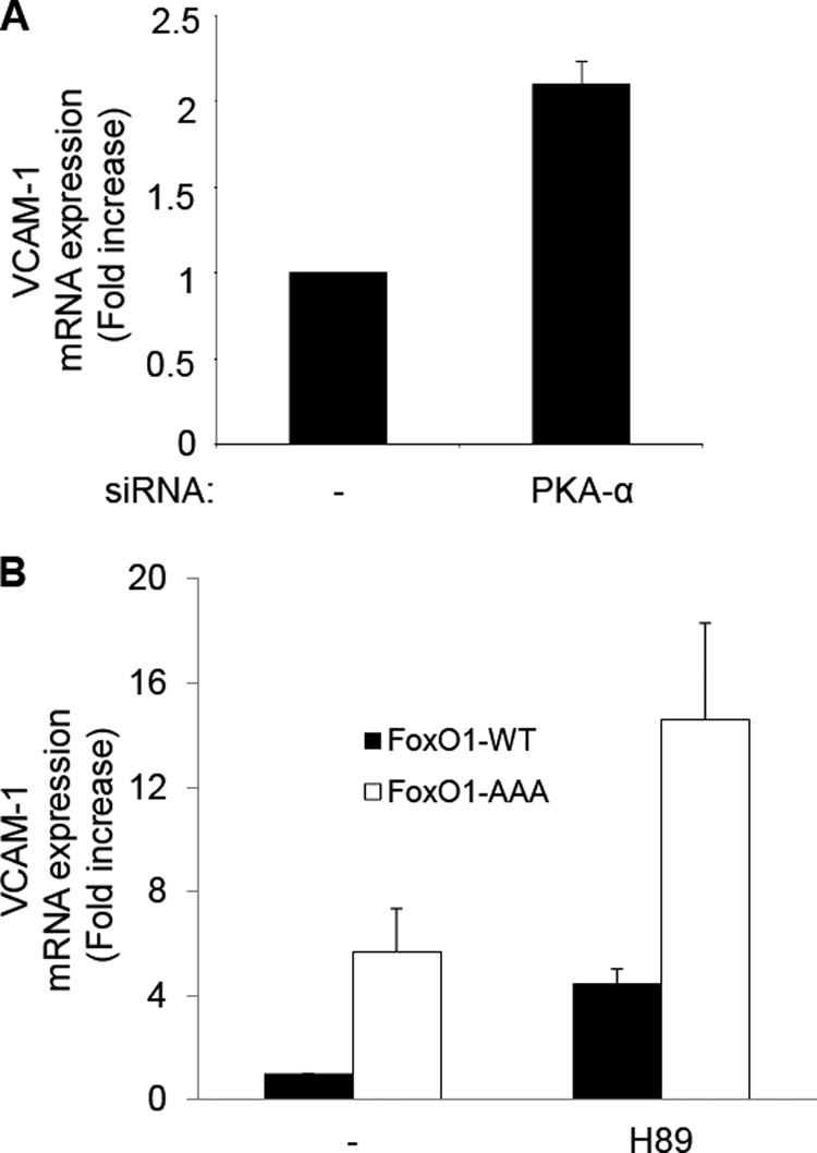
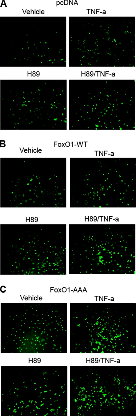
FoxO1, a forkhead box O class transcription factor, is abundant in insulin-responsive tissues. Akt, downstream from phosphatidylinositol 3-kinase in insulin signaling, phosphorylates FoxO1 at Thr(24), Ser(256), and Ser(319), negatively regulating its function. We previously reported that dehydroepiandrosterone-stimulated phosphorylation of FoxO1 in endothelial cells requires cAMP-dependent protein kinase α (PKA-α). Therefore, we hypothesized that FoxO1 is a novel direct substrate for PKA-α. Using an immune complex kinase assay with [γ-(32)P]ATP, purified PKA-α directly phosphorylated wild-type FoxO1 but not FoxO1-AAA (mutant with alanine substitutions at known Akt phosphorylation sites). Phosphorylation of wild-type FoxO1 (but not FoxO1-AAA) was detectable using phospho-specific antibodies. Similar results were obtained using purified GST-FoxO1 protein as the substrate. Thus, FoxO1 is a direct substrate for PKA-α in vitro. In bovine aortic endothelial cells, interaction between endogenous PKA-α and endogenous FoxO1 was detected by co-immunoprecipitation. In human aortic endothelial cells (HAEC), pretreatment with H89 (PKA inhibitor) or siRNA knockdown of PKA-α decreased forskolin- or prostaglandin E(2)-stimulated phosphorylation of FoxO1. In HAEC transfected with a FoxO-promoter luciferase reporter, co-expression of the catalytic domain of PKA-α, catalytically inactive mutant PKA-α, or siRNA against PKA-α caused corresponding increases or decreases in transactivation of the FoxO promoter. Expression of vascular cellular adhesion molecule-1 mRNA, up-regulated by FoxO1 in endothelial cells, was enhanced by siRNA knockdown of PKA-α or treatment of HAEC with the PKA inhibitor H89. Adhesion of monocytes to endothelial cells was enhanced by H89 treatment or overexpression of FoxO1-AAA, similar to effects of TNF-α treatment. We conclude that FoxO1 is a novel physiological substrate for PKA-α in vascular endothelial cells.


Figures

FIGURE 1.
FIGURE 1.
PKA-α directly phosphorylates FoxO1 in vitro. HEK293 cells were transfected with empty vector (pcDNA3) or expression vectors for FLAG-tagged FoxO1-WT or FoxO1-AAA. Two days after transfection, recombinant FoxO1 was immunoprecipitated from cell lysates (1 mg of total protein) using an anti-FLAG antibody. These recombinant FoxO1 proteins were used as the substrate along with purified PKA-α as the enzyme for an in vitro kinase assay as described under “Materials and Methods.” A, autoradiogram of a representative immune complex kinase assay is shown in the top panel. Samples from the kinase assay were immunoblotted (IB) using antibodies against FoxO1 (middle panel) or PKAα (bottom panel), demonstrating the appropriate absence or presence of the substrate (FoxO1) and the kinase (PKA-α). B, 32P-labeled FoxO1 from three independent experiments as shown in A was quantified using a Phospho-Imager and normalized to FoxO1 expression and is represented as the mean ± S.E. (32P-labeled FoxO1-WT in the presence of purified PKA-α is significantly greater that 32P-labeled FoxO1 in all other groups, p < 0.03; 32P-labeled FoxO1 in the absence of PKA-α is not statistically different from phosphorylated FoxO1-AAA in the presence of PKA-α, p > 0.7).
FIGURE 2.
FIGURE 2.
PKA-α specifically phosphorylates FoxO1 at Thr24, Ser256, and Ser319in vitro. Recombinant FoxO1-WT and FoxO1-AAA were obtained and subjected to an in vitro kinase assay with PKA-α as described in the legend to Fig. 1. A, samples were subjected to immunoblotting (IB) using antibodies that specifically detect phospho-FoxO1 at Thr24, Ser256, or Ser319. Samples from the kinase assay were also immunoblotted using antibodies against FoxO1 or PKAα to demonstrate the appropriate absence or presence of the substrate (FoxO1) and the kinase (PKA-α). Representative immunoblots are shown from a single experiment that was repeated independently four times. B, results from four independent experiments as shown in A were quantified using scanning densitometry and normalized to total FoxO1 expression and are represented as the means ± S.E. In the presence of PKA-α, wild-type FoxO1 (but not FoxO1-AAA) was significantly phosphorylated at Thr24 and Ser256 (when compared with all other corresponding groups, p < 0.01). Phosphorylation of wild-type FoxO1 at Ser319 in the presence of PKA-α was also significantly greater than that in the absence of PKA-α (compare the sixth and ninth bars; p < 0.05).
FIGURE 3.
FIGURE 3.
PKA-α directly phosphorylates purified GST-FoxO1 protein at Thr24, Ser256, and Ser319in vitro. Commercially obtained purified GST-FoxO1 protein or GST control protein (1 μg, as described under “Materials and Methods”) was used as the substrate along with purified PKA-α (0.1 μg) as the enzyme and for in vitro kinase assays as in Fig. 1. A, autoradiogram from a representative kinase assay using [γ-32P]ATP (top panel). Aliquots from each kinase assay were also immunoblotted using antibodies against FoxO1 (middle panel) or PKA-α (bottom panel), demonstrating the appropriate absence or presence of the substrate (GST-FoxO1) and the kinase (PKA-α). B, 32P-labeled GST-FoxO1 from three independent experiments as shown in A was quantified using a Phospho-Imager and normalized to GST-FoxO1 expression and is represented as the mean ± S.E. 32P-Labeled GST-FoxO1 in the presence of purified PKA-α is significantly greater than that in all other groups; p < 0.03). C, samples from in vitro kinase assays conducted without [γ-32P]ATP were subjected to immunoblotting using antibodies that specifically detect phospho-FoxO1 at Thr24, Ser256, or Ser319. Aliquots from each kinase assay were also immunoblotted using antibodies against FoxO1 or PKA-α to demonstrate the appropriate absence or presence of the substrate (GST-FoxO1) and the kinase (PKA-α). Representative immunoblots are shown from a single experiment that was repeated independently three times. D, results from three independent experiments as shown in C were quantified using scanning densitometry and normalized to total GST-FoxO1 expression and are represented as the means ± S.E. In the presence of PKA-α, GST-FoxO1 was significantly phosphorylated at Thr24, Ser256, and Ser319 (when compared with all other corresponding groups; p < 0.004).
FIGURE 4.
FIGURE 4.
Recombinant and endogenous PKA-α and FoxO1 interact with each other in intact cells. A, HEK293 cells were co-transfected with empty vector or FLAG-tagged FoxO1-WT in the absence or presence of HA-tagged PKA-α. Cell lysates from each group (1 mg total protein) were subjected to immunoprecipitation (IP) using anti-HA antibodies followed by immunoblotting (IB) using antibodies against FoxO1 or HA. Samples of whole cell lysates from the same experiments were also immunoblotted using antibodies against FLAG, HA, or β-actin to demonstrate the appropriate absence or presence of enzyme and substrate in each sample and comparable loading of samples. Representative immunoblots are shown for experiments that were repeated independently three times. B, total cell lysates prepared from nontransfected BAEC were subjected to immunoprecipitation using antibodies against PKA-α or FoxO1. The samples were also subjected to immunoprecipitation using nonimmune IgG as a negative control. Samples from each group were then subjected to immunoblotting using anti-FoxO1 antibodies. Total cell lysates (lane 4) were also immunoblotted with anti-FoxO1 antibodies to confirm the presence of endogenous FoxO1 (lane 4). Representative immunoblots are shown for experiments that were repeated independently three times.
FIGURE 5.
FIGURE 5.
Forskolin-stimulated phosphorylation of FoxO1 is prevented by pretreatment with PKA inhibitor H89 and by siRNA knockdown of PKA-α in HAEC. A, HAEC were serum-starved overnight and pretreated with vehicle or H89 (20 μm, 30 min) followed by treatment with vehicle or forskolin (20 μm, 30 min). Total cell lysates were subjected to immunoblotting using antibodies against phospho-FoxO1 (Ser256), FoxO1, or α-tubulin. Representative immunoblots are shown for experiments that were repeated independently four times. B, results from three independent experiments as shown in A were quantified using scanning densitometry and normalized to total FoxO1 expression and are represented as the means ± S.E. Forskolin treatment (second bar) significantly stimulated phosphorylation of FoxO1 (when compared with control (first bar), p < 0.01). This effect of forskolin was completely inhibited in cells pretreated with H89 (third bar versus first bar, p > 1.0). C, HAEC were transfected with nontargeting siRNA (control, lanes 1 and 3) or siRNA specifically targeting PKA-α (lanes 2 and 4) as described under “Materials and Methods.” 24 h after transfection, HAEC were serum-starved overnight and then treated with vehicle (Me2SO, lanes 1 and 2) or forskolin (20 μm, lanes 3 and 4) for 30 min. Total cell lysates from each group were subjected to immunoblotting using antibodies against phospho-FoxO1 (Ser256), PKA-α, FoxO1, or α-tubulin. Representative immunoblots are shown for experiments that were repeated independently four times. D, results from three independent experiments as shown in C were quantified by scanning densitometry. The amounts of phospho-FoxO1 were normalized to total FoxO1 (mean ± S.E.). For samples transfected with control siRNA, forskolin treatment significantly increased p-FoxO1 (Ser256) (p < 0.02). Phosphorylation of FoxO1 (Ser256) in cells transfected with siRNA targeting endogenous PKA-α in either the absence or presence of forskolin treatment was significantly decreased when compared with corresponding groups of cells transfected with control siRNA (p < 0.03).
FIGURE 6.
FIGURE 6.
PGE2-stimulated phosphorylation of FoxO1 is prevented by pretreatment with PKA inhibitor H89 and by siRNA knockdown of PKA-α in HAEC. A, HAEC were serum-starved overnight and pretreated with vehicle or H89 (20 μm, 30 min) followed by treatment with vehicle or PGE2 (500 nm, 30 min). Total cell lysates were subjected to immunoblotting using antibodies against phospho-FoxO1 (Ser256), FoxO1, or α-tubulin. Representative immunoblots are shown for experiments that were repeated independently three times. B, results from three independent experiments as shown in A were quantified using scanning densitometry and normalized to total FoxO1 expression and are represented as the means ± S.E. PGE2 treatment (second bar) significantly stimulated phosphorylation of FoxO1 (when compared with control (first bar), p < 0.03). This effect of PGE2 was completely inhibited in cells pretreated with H89 (third bar versus first bar, p > 0.9). C, HAEC were transfected with nontargeting siRNA (control, lane 1) or siRNA specifically targeting PKA-α (lanes 2 and 3) as described under “Materials and Methods.” 24 h after transfection, HAEC were serum-starved overnight and then treated with vehicle (lane 1) or PGE2 (500 nm, lanes 2 and 3) for 30 min. Total cell lysates from each group were subjected to immunoblotting using antibodies against phospho-FoxO1 (Ser256), PKA-α, FoxO1, or α-tubulin. Representative immunoblots are shown for experiments that were repeated independently three times. D, results from three independent experiments as shown in C were quantified by scanning densitometry. The amounts of phospho-FoxO1 were normalized to total FoxO1 (means ± S.E.). For samples transfected with control siRNA, PGE2 treatment significantly increased p-FoxO1 (Ser256) (p < 0.02). Phosphorylation of FoxO1 (Ser256) in cells transfected with siRNA targeting endogenous PKA-α in the presence of PGE2 treatment was significantly decreased when compared with PGE2-treated cells transfected with control siRNA (p < 0.03).
FIGURE 7.
FIGURE 7.
PKA-α regulates transcriptional activity of FoxO. A, HAEC were co-transfected with a FoxO promoter luciferase reporter and expression vectors for pcDNA (control), PKAα-cat-WT, or PKAα-cat-KD. Two days after transfection, the cell lysates were then subjected to dual-luciferase reporter assay as described under “Materials and Methods.” The data shown are the means ± S.E. of three independent experiments. Transcriptional activity of FoxO was substantially inhibited in cells transfected with PKA-α-cat-WT (when compared with the pcDNA control group, p < 0.03). By contrast, transcriptional activity of FoxO was substantially enhanced in cells transfected with PKA-α-cat-KD (p < 0.005). B, HAEC were co-transfected with nontargeting control siRNA or siRNA specifically targeting PKA-α along with the FoxO promoter luciferase reporter construct. Two days later, the lysates were subjected to a dual-luciferase reporter assay. The data shown are the means ± S.E. of three independent experiments. Transcriptional activity of FoxO was significantly increased by siRNA knockdown of endogenous PKA-α (when compared with control cells transfected with nontargeting siRNA; p < 0.05).
FIGURE 8.
FIGURE 8.
PKA-α regulates transcriptional activity of VCAM-1 through FoxO1. A, HAEC were transfected with nontargeting control siRNA (first bar) or siRNA specifically targeting PKA-α (second bar) as described under “Materials and Methods.” The data shown are the means ± S.E. of three independent experiments. When compared with the control group, the cells transfected with siRNA specifically targeting PKA-α caused a significant increase in expression of VCAM-1 mRNA (p < 0.02). B, HAEC were co-transfected with expression vectors for FoxO1-WT or FoxO1-AAA. 24 h after transfection, HAEC were serum-starved overnight and then treated with vehicle (Me2SO, first and second bars) or H89 (10 μm, third and fourth bars) for 6 h. 1 μg of total RNA was reverse-transcribed and subjected to quantitative real time PCR for VCAM-1 and β-actin using QuantiTect SYBR Green PCR with appropriate primer sets as described under “Materials and Methods.” VCAM-1 mRNA expression was normalized to β-actin mRNA expression. The data shown are the means ± S.E. of four independent experiments. In both the absence and presence of H89, cells transfected with FoxO1-AAA significantly increased expression of VCAM-1 mRNA (compare first and second bars or third and fourth bars; p < 0.03). Moreover, in cells co-transfected with FoxO1-WT, treatment with H89 significantly enhanced expression of VCAM-1 mRNA (compare first and third bars; p < 0.001). Finally, in cells transfected with FoxO1-AAA, H89 treatment caused a significant increase in VCAM-1 mRNA (compare second and fourth bars; p < 0.02).
FIGURE 9.
FIGURE 9.
Overexpression of FoxO1 in human endothelial cells regulates TNF-α-stimulated expression of VCAM-1 and monocyte adhesion. HAEC were co-transfected with expression vectors for pcDNA (A), FoxO1-WT (B), or FoxO1-AAA (C). 24 h after transfection, HAEC were serum-starved for 1 h and then treated with vehicle, TNF-α (10 ng/ml), or H89 (20 μm) for 5 h. Calcein-AM-labeled U937 cells (6 × 10⁵) were incubated for 30 min at 37 °C with confluent HAEC. Chambered wells were then washed three times with PBS, and the cells were fixed in 2% formaldehyde. Labeled monocytes adhering to HAEC were visualized using an epifluorescent microscope as described under “Materials and Methods.” B, overexpression of FoxO1-WT and treatment with H89 (PKA-α inhibitor) tended to increase adhesion of monocytes to endothelium when compared with the same treatment in cells transfected with control pcDNA. C, moreover, overexpression of FoxO1-AAA and treatment of PKA-α inhibitor H89 tended to increase adhesion of monocytes to endothelium when compared with control pcDNA. In the absence of TNF-α treatment, when compared with vehicle-treated control cells, TNF-α treatment tended to cause increased adhesion of monocytes to endothelium.

Similar articles

Cited by

References

    1. Nakae J., Biggs W. H., 3rd, Kitamura T., Cavenee W. K., Wright C. V., Arden K. C., Accili D. (2002) Nat. Genet. 32, 245–253 - PubMed
    1. Accili D., Arden K. C. (2004) Cell 117, 421–426 - PubMed
    1. Chen H., Lin A. S., Li Y., Reiter C. E., Ver M. R., Quon M. J. (2008) J. Biol. Chem. 283, 29228–29238 - PMC - PubMed
    1. Furuyama T., Kitayama K., Shimoda Y., Ogawa M., Sone K., Yoshida-Araki K., Hisatsune H., Nishikawa S., Nakayama K., Nakayama K., Ikeda K., Motoyama N., Mori N. (2004) J. Biol. Chem. 279, 34741–34749 - PubMed
    1. Potente M., Urbich C., Sasaki K., Hofmann W. K., Heeschen C., Aicher A., Kollipara R., DePinho R. A., Zeiher A. M., Dimmeler S. (2005) J. Clin. Invest. 115, 2382–2392 - PMC - PubMed

Publication types

MeSH terms

Substances