. 2011 Mar 4;286(9):7619-28.
doi: 10.1074/jbc.M110.158477. Epub 2010 Dec 22.

N-cadherin regulates p38 MAPK signaling via association with JNK-associated leucine zipper protein: implications for neurodegeneration in Alzheimer disease

Affiliations

N-cadherin regulates p38 MAPK signaling via association with JNK-associated leucine zipper protein: implications for neurodegeneration in Alzheimer disease

Koichi Ando et al. J Biol Chem. .

Abstract

Synaptic loss, which strongly correlates with the decline of cognitive function, is one of the pathological hallmarks of Alzheimer disease. N-cadherin is a cell adhesion molecule essential for synaptic contact and is involved in the intracellular signaling pathway at the synapse. Here we report that the functional disruption of N-cadherin-mediated cell contact activated p38 MAPK in murine primary neurons, followed by neuronal death. We further observed that treatment with Aβ(42) decreased cellular N-cadherin expression through NMDA receptors accompanied by increased phosphorylation of both p38 MAPK and Tau in murine primary neurons. Moreover, expression levels of phosphorylated p38 MAPK were negatively correlated with that of N-cadherin in human brains. Proteomic analysis of human brains identified a novel interaction between N-cadherin and JNK-associated leucine zipper protein (JLP), a scaffolding protein involved in the p38 MAPK signaling pathway. We demonstrated that N-cadherin expression had an inhibitory effect on JLP-mediated p38 MAPK signal activation by decreasing the interaction between JLP and p38 MAPK in COS7 cells. Also, this study demonstrated a novel physical and functional association between N-cadherin and p38 MAPK and suggested neuroprotective roles of cadherin-based synaptic contact. The dissociation of N-cadherin-mediated synaptic contact by Aβ may underlie the pathological basis of neurodegeneration such as neuronal death, synaptic loss, and Tau phosphorylation in Alzheimer disease brain.


Figures
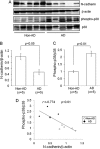
FIGURE 1.
FIGURE 1.
Increased phospho-p38 MAPK expressions were negatively correlated with decreased N-cadherin expressions in human brains. A, brain homogenates of temporal cortex from AD patients (AD, n = 5) and age-matched non-AD controls (Non-AD, n = 5) were analyzed by Western blot using anti-N-cadherin, β-actin, phospho-p38, and p38 MAPK antibodies. B, the band densities of N-cadherin and control β-actin were quantified by National Institutes of Health Image. The ratio of N-cadherin to β-actin (N-cadherin/β-actin) was calculated and analyzed by Mann-Whitney's U test. The N-cadherin/β-actin ratio was significantly decreased in the brains of AD patients compared with that of non-AD controls (p < 0.05). C, the band densities of phospho-p38 and p38 MAPK were quantified by National Institutes of Health Image. The phospho/total p38 MAPK ratio was calculated and analyzed by Mann-Whitney's U test. The phospho/total p38 MAPK ratio was significantly increased in the brains of AD patients compared with that of non-AD controls (p < 0.01). D, significant correlation was established in comparison between the phospho/total p38 MAPK ratio and the N-cadherin/β-actin ratio by Pearson's correlation co-efficients. The phospho/total p38 MAPK ratio was negatively correlated with the N-cadherin/β-actin ratio in human brain samples. (r = −0.774, p < 0.01).
FIGURE 2.
FIGURE 2.
ADH-1 induced neuronal cell death by activating p38 MAPK in murine primary neurons. A, ADH-1, N-cadherin antagonist, was applied to murine primary neurons at different concentrations as shown. 24 h after treatment, the lysates were immunoblotted with anti-phospho-p38 and p38 MAPK antibodies. ADH-1 increased phosphorylation of p38 MAPK. B, neuronal cell death induced by ADH-1 was evaluated by MTT assay (n = 5, p < 0.001). C, murine primary neurons were treated with ADH-1 (0.5 mg/ml) for 24 h with or without 30 min of pretreatment with 10 μm SB203580, a specific p38 MAPK inhibitor, and the cell death was examined by MTT assay (n = 4, p < 0.001). SB203580 attenuated neuronal cell death induced by ADH-1 (n = 4, p < 0.001). D, ADH-1 (0.5 mg/ml) was added to confluent SH-SY5Y cells for 24 h followed by the immunostaining, using anti-N-cadherin and anti-phospho-p38 antibodies. Treatment with ADH-1 perturbed N-cadherin immunoreactivity, indicating a partial loss of N-cadherin from cell-cell junctions. Scale bar, 10 μm.
FIGURE 3.
FIGURE 3.
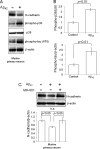
42 decreased N-cadherin expressions through NMDA receptors in murine primary neurons. A, murine primary neurons were treated with or without 100 nm synthetic Aβ42 peptides for 48 h, and the lysates were immunoblotted with anti-N-cadherin, phospho-p38, p38 MAPK, phospho-Tau (AT8), and β-actin antibodies, successively. Aβ42 treatment reduced N-cadherin expressions and induced phosphorylation of p38 MAPK and Tau in murine primary neurons. B, the band densities of phospho-p38, p38 MAPK, phospho-Tau (AT8), and β-actin were quantified by National Institutes of Health Image. The ratios of phospho/total p38 MAPK and phospho-Tau (AT8)/β-actin were calculated and analyzed by Student's t test (n = 3, p < 0.01). C, murine primary neurons were pretreated with 10 μm MK-801, NMDA receptor antagonist for 30 min followed by 100 nm synthetic Aβ42 peptides for 48 h. The lysates were evaluated by Western blot using anti-N-cadherin and β-actin antibodies. The band densities of N-cadherin and β-actin were quantified by National Institutes of Health Image. The N-cadherin/β-actin ratio was calculated and analyzed by one-way ANOVA. NMDA receptor antagonist MK-801 prevented the Aβ42-induced decrease in N-cadherin levels (n = 3, p < 0.05).
FIGURE 4.
FIGURE 4.
The association of N-cadherin with JLP in HEK293 cells and neuronal cells. A, lysates of HEK293 cells, transiently transfected with HA-tagged N-cadherin and/or FLAG-tagged JLP expressing vectors, were immunoprecipitated (IP) with anti-HA antibody (lanes 1–3) or normal IgG (lane 4). The immunoprecipitates and the lysates were analyzed by immunoblotting (IB) with the specific antibodies against the FLAG tag and HA tag. B, lysates of HEK293 cells, transiently transfected with HA-tagged N-cadherin and/or FLAG-tagged JLP expressing vectors were immunoprecipitated with anti-FLAG antibody (lanes 1–3) or normal IgG (lane 4). The immunoprecipitates and the lysates were analyzed by Western blot with the specific antibodies against the HA tag and FLAG tag. C, the endogenous association between N-cadherin and JLP in murine primary neurons was analyzed. The neurons were lysed and immunoprecipitated with either anti-N-cadherin antibody or normal IgG. The immunoprecipitates and the lysates were immunoblotted with the specific antibodies against JLP and N-cadherin.
FIGURE 5.
FIGURE 5.
Deletion mapping of N-cadherin-interacting domain of JLP. A, schematic diagram of the C-terminally truncated S-tagged JLP constructs used for analysis of the JLP-N-cadherin interaction. The sizes of these deletion mutants were 374 (amino acids 1–374), 467 (amino acids 1–467), 1000 (amino acids 1–1000), 1165 (amino acids 1–1165), and 1307 amino acids (full length, residues 1–1307), respectively. B, COS7 cells were transiently transfected with the WT or C-terminally truncated mutants of S-tagged JLP. The lysates were immunoprecipitated (IP) with anti-N-cadherin antibody. The immunoprecipitates and the lysates were subjected to Western blot analysis with the specific antibodies against S-tag. All of these deletion mutants of JLP were co-immunoprecipitated with N-cadherin. C, schematic diagram representing shorter constructs of C-terminally truncated S-tagged JLP. N-terminally truncated short constructs were also prepared as shown in Fig. 5C. D, COS7 cells were transiently transfected with these different mutants of S-tagged JLP. The lysates were immunoprecipitated with anti-N-cadherin antibody, and the immunoprecipitates were immunoblotted (IB) with anti-S-tag antibody. The mutants consisting of amino acids 160–398 and 160–463 of JLP were co-immunoprecipitated with N-cadherin, whereas the mutant consisting of amino acids 210–398 of JLP was not co-immunoprecipitated with N-cadherin.
FIGURE 6.
FIGURE 6.
N-cadherin expression inhibited JLP-associated p38 MAPK pathway. A, COS7 cells were co-transfected with FLAG-tagged p38α, FLAG-tagged MKK4, S-tagged JLP, and HA-tagged N-cadherin. 24 h after transfection, the lysates were precipitated with S-protein-agarose. The lysates and precipitates were subjected to Western blot analysis with anti-phospho-p38 or p38 MAPK antibodies. One representative immunoblot result is shown. The band densities of phospho-p38 and p38 MAPK were quantified by National Institutes of Health Image. The phospho/total p38 MAPK ratio was calculated and analyzed by one-way ANOVA. N-cadherin expression significantly inhibited JLP/MKK4-mediated phosphorylation of p38 MAPK compared with that without expression of N-cadherin (n = 3, p < 0.05). Pull-down assay using S-protein-agarose showed that N-cadherin expression decreased the interaction between JLP and p38 MAPK. B, COS7 cells were co-transfected with FLAG-tagged p38α, HA-tagged MEKK3, S-tagged JLP, and HA-tagged N-cadherin. 24 h after transfection, the lysates were subjected to Western blot analysis with anti-phospho-p38 or p38 MAPK antibodies. One representative immunoblot result is shown. The band densities of phospho-p38 and p38 were quantified by National Institutes of Health Image. The ratio of phospho/total p38 MAPK was calculated and analyzed by one-way ANOVA. N-cadherin expression inhibited JLP/MEKK3-mediated phosphorylation of p38 MAPK significantly compared with that without expression of N-cadherin (n = 3, p < 0.05). Pull-down assay using S-protein-agarose showed that N-cadherin expression inhibited the interaction between JLP and p38 MAPK.
FIGURE 7.
FIGURE 7.
Hypothetical model of AD pathogenesis caused by the disruption of N-cadherin-based synaptic contact. Left panel, in the basal state at the synapse, N-cadherin-based synaptic adhesion stabilizes JLP, which suppresses neurotoxic p38 MAPK activation. Right panel, in AD, when N-cadherin-based synaptic contact is disrupted by Aβ42, Aβ42 could decrease N-cadherin expressions resulting in aberrant p38 MAPK activation followed by the subsequent Tau phosphorylation and neuronal death.

References

    1. Terry R. D., Masliah E., Salmon D. P., Butters N., DeTeresa R., Hill R., Hansen L. A., Katzman R. (1991) Ann. Neurol. 30, 572–580 - PubMed
    1. Selkoe D. J. (2002) Science 298, 789–791 - PubMed
    1. Shankar G. M., Bloodgood B. L., Townsend M., Walsh D. M., Selkoe D. J., Sabatini B. L. (2007) J. Neurosci. 27, 2866–2875 - PMC - PubMed
    1. Wolfe M. S., De Los Angeles J., Miller D. D., Xia W., Selkoe D. J. (1999) Biochemistry 38, 11223–11230 - PubMed
    1. Wolfe M. S., Xia W., Ostaszewski B. L., Diehl T. S., Kimberly W. T., Selkoe D. J. (1999) Nature 398, 513–517 - PubMed

MeSH terms