. 2011 Sep;45(3):550-6.
doi: 10.1165/rcmb.2010-0376OC. Epub 2010 Dec 22.

Fatty acid-binding proteins and peribronchial angiogenesis in bronchopulmonary dysplasia

Affiliations

Fatty acid-binding proteins and peribronchial angiogenesis in bronchopulmonary dysplasia

Elisa Ghelfi et al. Am J Respir Cell Mol Biol. 2011 Sep.

Abstract

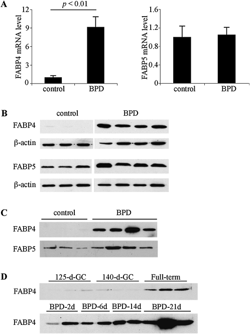
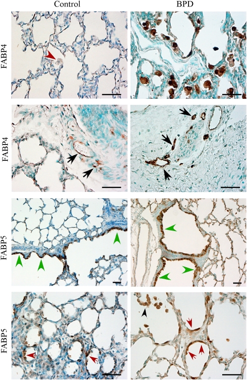
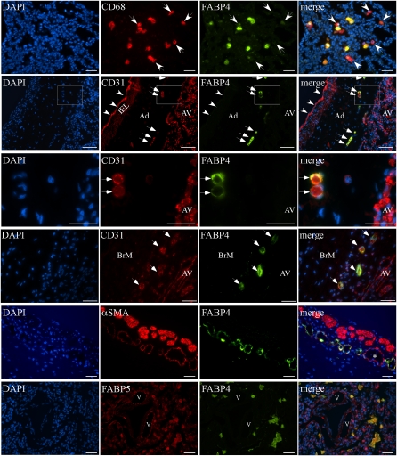
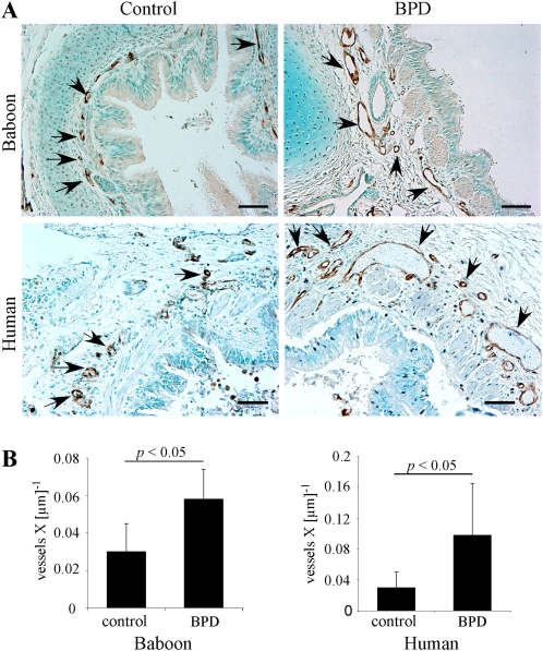
Inflammation plays a key role in the pathogenesis of bronchopulmonary dysplasia (BPD). Fatty acid-binding proteins (FABPs) 4 and 5 regulate the inflammatory activity of macrophages. Whether FABPs 4 and 5 could play a role in the pathogenesis of BPD via the promotion of macrophage inflammatory activity is unknown. This study sought to examine whether the expression levels of FABP4 and FABP5 were altered in bronchoalveolar lavage fluid and lung tissue in a baboon model of BPD. This study also sought to characterize the cell types that express these proteins. Real-time PCR, immunoblotting, immunohistochemistry, and double immunofluorescence were used to examine the expression of FABPs in samples of BPD. Morphometric analysis was used to quantify FABP4-positive peribronchial blood vessels in lung sections. FABP4 was primarily expressed in macrophages in samples of BPD. In addition, FABP4 was expressed in the endothelial cells of blood vessels in peribronchial areas and the vasa vasorum, but not in the alveolar vasculature in samples of BPD. FABP4 concentrations were significantly increased in lungs and bronchoalveolar lavage fluid samples with BPD. An increased density of FABP4-positive peribronchial blood vessels was evident in both baboon and human BPD sections. FABP5 was expressed in several cell types, including alveolar epithelial cells and macrophages. FABP5 concentrations did not show any significant alterations in BPD. In conclusion, FABP4 but not FABP5 levels are increased in BPD. FABP4 is differentially expressed in endothelial cells of the bronchial microvasculature, which demonstrates a previously unrecognized expansion in BPD.


Figures

Figure 1.
Figure 1.
The expression of fatty acid–binding protein 4 (FABP4) is increased in bronchopulmonary dysplasia (BPD). (A) Real-time RT-PCR for FABP4 and FABP5 was performed on total lung RNA, isolated from control and BPD group baboons (n = 6–7 samples per group). Relative expression levels were normalized to β-actin by the 2−ΔΔCT method. An arbitrary level of 1 was assigned to the control group. (B) FABP4 and FABP5 protein expression in whole baboon lung homogenates was analyzed by immunoblotting. β-actin was used as a loading control. (C) FABP4 and FABP5 were detected by immunoblotting in necropsy bronchoalveolar lavage fluid (BALF). (D) Time course of FABP4 detection in necropsy BALF samples.
Figure 2.
Figure 2.
Immunolocalization of FABP4 and FABP5 in a baboon model of BPD. Representative images are shown. FABP4 was detected in macrophages in BPD, but not in control samples (first row, red arrowhead). FABP4 was also detected in peribronchial vascular endothelial cells in both control and BPD samples (second row, black arrowheads). FABP5 was localized to bronchiolar (third row, green arrowheads) and alveolar epithelial Type 2 cells (fourth row, red arrowheads), as well as macrophages (fourth row, black arrowhead). Scale bars = 50 μm.
Figure 3.
Figure 3.
Double immunofluorescence analysis for FABPs, CD31, CD68, and α-smooth muscle actin (αSMA) on baboon and human BPD sections. Representative images are shown. First row (baboon BPD): FABP4 is colocalized with CD68 in most, but not all, alveolar macrophages. White arrows indicate CD68-positive, but FABP4-negative, cells. Second row (human BPD): CD31 and FABP4 are colocalized in vasa vasorum ECs in the adventitia (Ad) of a pulmonary artery (white arrows), but not in pulmonary arterial ECs (white arrowheads) or alveolar vessels (AV). Internal elastic lamina (IEL) demonstrates autofluorescence. Third row: Higher magnification of the boxed area in row 2 demonstrates CD31 expression in alveolar vasculature and vasa vasorum ECs, but FABP4 expression only in vasa vasorum ECs (white arrows). Fourth row (human BPD): CD31 and FABP4 are colocalized in small vascular ECs in the bronchial mucosa (BrM, white arrows). Fifth row (human BPD): αSMA, a marker of mature pericytes and smooth muscle cells, is detected around some FABP4-positive peribronchial vessels. Larger vessels with continuous coverage of αSMA-expressing cells do not express FABP4 (asterisk). Sixth row (human BPD): In contrast to the widespread expression pattern of FABP5 in several cell types, including alveolar epithelial and endothelial cells lining pulmonary vessels (v) in the lung parenchyma, FABP4 is detected only in FABP5-positive alveolar macrophages. Scale bars = 25 μm.
Figure 4.
Figure 4.
FABP4-positive peribronchial blood vessel density is increased in BPD. (A) Representative images of immunohistochemistry for FABP4 demonstrate FABP4-positive peribronchial vessels (black arrowheads) in baboon and human lungs with and without BPD. Scale bars = 50 μm. (B) Peribronchial blood vessel density was quantitated and normalized to the airway perimeter in baboon and human infants with BPD (n = 5 samples per group).

References

    1. Fanaroff AA, Stoll BJ, Wright LL, Carlo WA, Ehrenkranz RA, Stark AR, Bauer CR, Donovan EF, Korones SB, Laptook AR, et al. 2007. Trends in neonatal morbidity and mortality for very low birthweight infants. Am J Obstet Gynecol 2007;147.e1–8 - PubMed
    1. Baraldi E, Filippone M. Chronic lung disease after premature birth. N Engl J Med 2007;357:1946–1955 - PubMed
    1. Eichenwald EC, Stark AR. Management and outcomes of very low birth weight. N Engl J Med 2008;358:1700–1711 - PubMed
    1. Kallapur SG, Jobe AH. Contribution of inflammation to lung injury and development. Arch Dis Child Fetal Neonatal Ed 2006;91:F132–F135 - PMC - PubMed
    1. Bancalari E, Claure N. Non-invasive ventilation of the preterm infant. Early Hum Dev 2008;84:815–819 - PubMed

Publication types

Substances