. 2011 Mar;105(3):1033-44.
doi: 10.1152/jn.00810.2010. Epub 2010 Dec 22.

Alterations of action potentials and the localization of Nav1.6 sodium channels in spared axons after hemisection injury of the spinal cord in adult rats

Affiliations

Alterations of action potentials and the localization of Nav1.6 sodium channels in spared axons after hemisection injury of the spinal cord in adult rats

Arsen S Hunanyan et al. J Neurophysiol. 2011 Mar.

Abstract

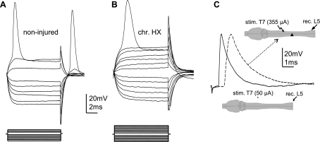
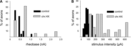
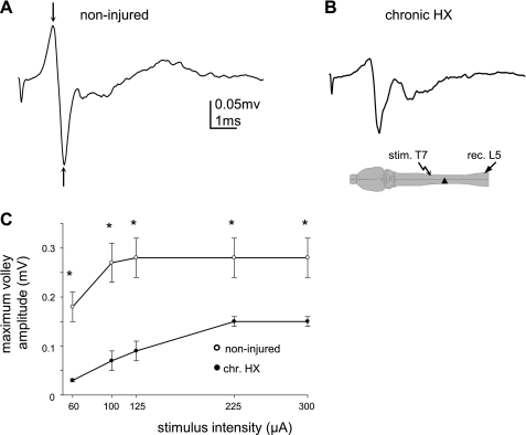
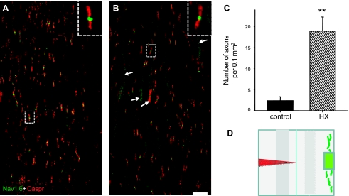
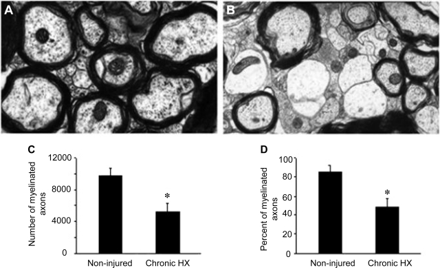
Previously, we reported a pronounced reduction in transmission through surviving axons contralateral to chronic hemisection (HX) of adult rat spinal cord. To examine the cellular and molecular mechanisms responsible for this diminished transmission, we recorded intracellularly from lumbar lateral white matter axons in deeply anesthetized adult rats in vivo and measured the propagation of action potentials (APs) through rubrospinal/reticulospinal tract (RST/RtST) axons contralateral to chronic HX at T10. We found decreased excitability in these axons, manifested by an increased rheobase to trigger APs and longer latency for AP propagation passing the injury level, without significant differences in axonal resting membrane potential and input resistance. These electrophysiological changes were associated with altered spatial localization of Nav1.6 sodium channels along axons: a subset of axons contralateral to the injury exhibited a diffuse localization (>10 μm spread) of Nav1.6 channels, a pattern characteristic of demyelinated axons (Craner MJ, Newcombe J, Black JA, Hartle C, Cuzner ML, Waxman SG. Proc Natl Acad Sci USA 101: 8168-8173, 2004b). This result was substantiated by ultrastructural changes seen with electron microscopy, in which an increased number of large-caliber, demyelinated RST axons were found contralateral to the chronic HX. Therefore, an increased rheobase, pathological changes in the distribution of Nav1.6 sodium channels, and the demyelination of contralateral RST axons are likely responsible for their decreased conduction chronically after HX and thus may provide novel targets for strategies to improve function following incomplete spinal cord injury.


Figures

Fig. 1.
Fig. 1.
Comparison of the action potentials (APs) measured intracellularly from an L5 motoneuron (MN) and intra-axonally from a lateral white matter axon and a dorsal root (DR) axon in the spinal cord of the same adult rat. A: intracellular recording from the L5 MN in response to electrical stimulation of the lateral white matter ipsilaterally at T7 with rising stimulus intensity; the intensity of electrical stimulation was 30 μA [1, dotted trace; stimulus intensity was at maximum to evoke the maximum excitatory postsynaptic potential (EPSP) response] and 80 μA (2, solid trace; stimulus intensity was the minimum required to evoke an AP), respectively. B: intra-axonal recording from a L5 rubrospinal tract (RST) axon in response to electrical stimulation of the lateral white matter ipsilaterally at T7; the intensity of electrical stimulation was 80 μA (1) and 90 μA (2; minimum stimulus intensity required to trigger AP in this particular axon), respectively. C: intra-axonal recording from a L5 dorsal root axon in response to electrical stimulation of the fasciculus gracilis at T7; the intensity of electrical stimulation was 140 μA (1) and 150 μA (2; minimum stimulus intensity required to trigger AP), respectively. Diagrams below the traces show the positions of the stimulating (st.) and recording (rec.) electrodes. Note the EPSP phase is before the AP when recorded from MNs, but there is an absence of such an EPSP phase when the recording is made either from the lateral white matter or from DR axons.
Fig. 2.
Fig. 2.
Comparison of the pharmacological properties of EPSPs recorded from L5 MNs vs. APs recorded from L5 lateral white matter axons, both evoked by electrical stimulation of ipsilateral white matter at T7. A–C: EPSP responses from a MN before (A) and after 6-cyano-7-nitroquino-xaline-2,3-dione (CNQX) injections into the gray matter close to the recording electrode (B) and after tetrodotoxin (TTX) injections into the white matter rostral/ipsilateral to the recording electrode (C). D–F: axonal AP before (D) and after CNQX injections close and rostral to the recording electrode (E) and after TTX injections into the white matter rostral/ipsilateral to the recording electrode (F). Note that CNQX injections blocked EPSPs in the MN but did not affect the propagation of an axonal AP. Further TTX injection into the white matter blocked the axonal AP.
Fig. 3.
Fig. 3.
Intra-axonal recording from the lateral white matter axons was done to demonstrate the decreased excitability (higher rheobase current), decreased conduction velocity (increased latency), and absence of changes in the resting membrane potential (RMP) in RST axons passing across from chronic hemisection (chr. HX). Changes in the properties of APs recorded intra-axonally from RST axons contralateral to the chronic HX spinal cord injury (SCI) are shown. A and B: representative traces show voltage membrane responses in single axons to current steps passed through the recording electrode in noninjured (A) and chronic HX SCI rats (B). Both axons had a RMP of −60 mV. Current steps (displayed below the voltage traces) of a 0.1-nA increment were applied through the recording electrode in both hyperpolarizing (to measure membrane resistance) and depolarizing directions (to trigger an AP). Note the higher rheobase but similar membrane resistance in the axon from chronic HX spinal cord. C: superimposed examples of APs in normal (solid trace) and chronic HX SCI rats (dashed trace) to demonstrate a longer latency of the AP recorded from L5 lateral white matter axons in response to electrical stimulation of T7 ipsilateral white matter. Both axons displayed a RMP of −63 mV. Diagram shows the positions of the recording (rec.) and stimulating (stim.) electrodes in normal (bottom) and chronic HX cord (arrow, top).
Fig. 4.
Fig. 4.
Histograms show the difference in the electrical properties of single axons recorded intra-axonally within the L5 lateral white matter in noninjured and chronic HX SCI rats. A: the rheobase current at the recording electrode required to trigger an AP. B: the minimum stimulus intensity at the T7 stimulating electrode required to evoke an AP in L5 lateral white matter axons. Note an increased percentage of axons that have a relatively high rheobase (A) and require a relatively high stimulus intensity to evoke an AP (B) in HX SCI rats. For comparison of the noninjured control and chronic HX SCI animals, we used axons that displayed a similar RMP.
Fig. 5.
Fig. 5.
Diminished extracellular responses recorded from the L5 lateral white matter and evoked by electrical stimulation of the lateral white matter ipsilaterally at T7 in chronic HX SCI rats. A and B: representative traces of the volley of APs in noninjured (A) and chronic HX SCI rats (B). Diagram shows the configuration of the stimulation and recording electrodes. C: summary of results indicating the diminished maximum volley responses and the higher stimulus intensity required to induce these responses in chronic HX SCI rats. *P < 0.05.
Fig. 6.
Fig. 6.
Double immunostaining with Nav1.6 and Caspr to show their axonal localization at the T10 level on right lateral white matter of noninjured rats and left lateral white matter of chronic HX SCI rats. A: normal spinal cord. Note the expression of Nav1.6 was found mainly within the nodes of Ranvier. B: a diffuse localization pattern of Nav1.6 channels was observed after chronic HX SCI. Small arrows point to “wormlike” axonal profiles with diffuse (>10 μm) Nav1.6 sodium channel immunostaining. Large arrow shows the localization of Nav1.6 channels within the nodes of Ranvier on an axon also with diffuse Nav1.6 sodium channel expression. Insets in A and B are micrographs showing sodium channel immunostaining within the nodes at 10 times higher magnification. Scale bar, 20 μm. C: the number of axons with a diffuse localization of Nav1.6 in normal (n = 3) and chronic HX SCI rats (n = 3). Data are means ± SE. **P < 0.01. D: the highlighted area contralateral to the HX SCI on the diagram represents the region from which the images in A and B were taken.
Fig. 7.
Fig. 7.
Number and percentage of myelinated axons in an area corresponding to the RST within the contralateral white matter to the HX SCI shows that they are reduced in number during the chronic stage of injury. Compared with noninjured controls (A), there were significantly fewer myelinated axons at 6 wk postinjury (B). C and D: quantification of the numbers of myelinated axons (C) and the percentage of myelinated axons (D) within the region corresponding to the RST in noninjured controls (n = 3) vs. 6 wk post-HX SCI rats (n = 6). Data are means ± SE. *P < 0.05.

Similar articles

Cited by

References

    1. Arvanian VL, Mendell LM. Removal of NMDA receptor Mg2+ block extends the action of NT-3 on synaptic transmission in neonatal rat motoneurons. J Neurophysiol 86: 123–129, 2001 - PubMed
    1. Arvanian VL, Schnell L, Lou L, Golshani R, Hunanyan A, Ghosh A, Pearse DD, Robinson JK, Schwab ME, Fawcett JW, Mendell LM. Chronic spinal hemisection in rats induces a progressive decline in transmission in uninjured fibers to motoneurons. Exp Neurol 216: 471–480, 2009 - PMC - PubMed
    1. Baker M, Bostock H, Grafe P, Martius P. Function and distribution of three types of rectifying channel in rat spinal root myelinated axons. J Physiol 383: 45–67, 1987 - PMC - PubMed
    1. Ballermann M, Fouad K. Spontaneous locomotor recovery in spinal cord injured rats is accompanied by anatomical plasticity of reticulospinal fibers. Eur J Neurosci 23: 1988–1996, 2006 - PubMed
    1. Barrett EF, Barrett JN. Intracellular recording from vertebrate myelinated axons: mechanism of the depolarizing afterpotential. J Physiol 323: 117–144, 1982 - PMC - PubMed

Publication types

Substances