. 2011 Feb 1;186(3):1442-9.
doi: 10.4049/jimmunol.1003023. Epub 2010 Dec 22.

Transplantation tolerance to a single noninherited MHC class I maternal alloantigen studied in a TCR-transgenic mouse model

Affiliations

Transplantation tolerance to a single noninherited MHC class I maternal alloantigen studied in a TCR-transgenic mouse model

Yoshinobu Akiyama et al. J Immunol. .

Abstract

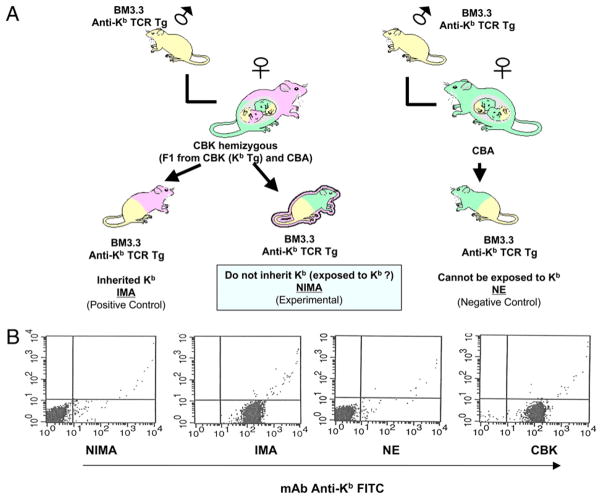
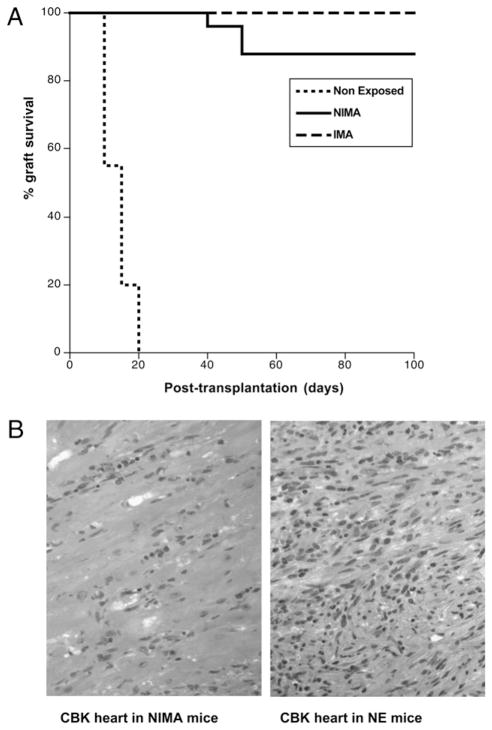
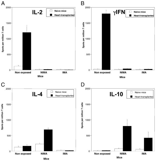
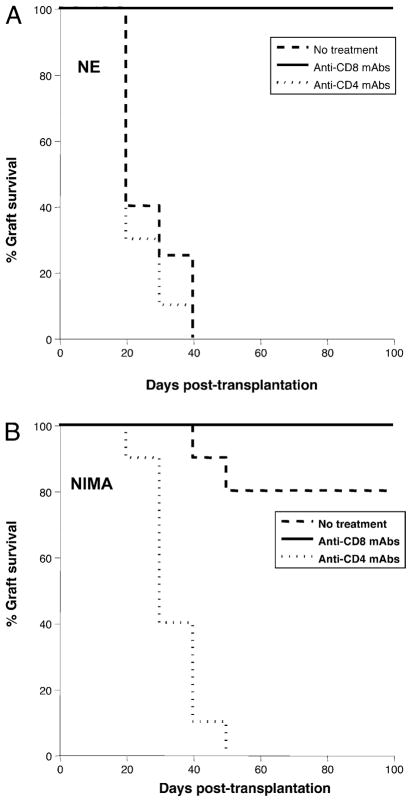
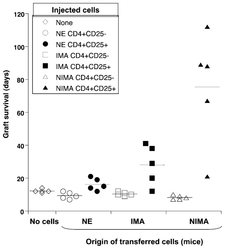
The mechanisms underlying tolerance to noninherited maternal Ags (NIMA) are not fully understood. In this study, we designed a double-transgenic model in which all the offspring's CD8(+) T cells corresponded to a single clone recognizing the K(b) MHC class I protein. In contrast, the mother and the father of the offspring differed by the expression of a single Ag, K(b), that served as NIMA. We investigated the influence of NIMA exposure on the offspring thymic T cell selection during ontogeny and on its peripheral T cell response during adulthood. We observed that anti-K(b) thymocytes were exposed to NIMA and became activated during fetal life but were not deleted. Strikingly, adult mice exposed to NIMA accepted permanently K(b+) heart allografts despite the presence of normal levels of anti-K(b) TCR transgenic T cells. Transplant tolerance was associated with a lack of a proinflammatory alloreactive T cell response and an activation/expansion of T cells producing IL-4 and IL-10. In addition, we observed that tolerance to NIMA K(b) was abrogated via depletion of CD4(+) but not CD8(+) T cells and could be transferred to naive nonexposed mice via adoptive transfer of CD4(+)CD25(high) T cell expressing Foxp3 isolated from NIMA mice.


Conflict of interest statement

Disclosures

The authors have no financial conflicts of interest.

Figures

FIGURE 1
FIGURE 1
The model. A, To obtain NIMA and IMA mice, we mated Bm3.3 anti-Kb TCR Tg male mice with (CBK [CBA, Kb Tg] × CBA) F1 female mice. The offspring, which inherited both the anti-Kb TCR transgene and the Kb transgene, were referred to as IMA mice. The offspring, which inherited the anti-Kb TCR transgene but not the Kb trans-gene, were referred to as NIMA mice. NE control offspring were obtained by mating BM3.3 anti-Kb TCR Tg female mice with CBA male mice (these mice could never be exposed to Kb). B, The expression of MHC class I glycoprotein Kb was assessed in positive control mice IMA and CBK, negative control mice NE, and experimental NIMA mice. Representative FACS profiles obtained with spleen cells are shown.
FIGURE 2
FIGURE 2
NIMA mice accept Kb+ allogeneic CBK heart transplants. NE, IMA (express Kb), and NIMA (experimental group) mice underwent het-erotopic vascularized transplantation with a heart derived from a CBK (Kb Tg) mouse. Transplant rejection was monitored for 100 d by palpation (heartbeat) and histological methods. A, Percentages of graft survival at different time points after transplantation. Graft survival was analyzed using the Kaplan–Meier method, and survival curves were compared using the log-rank test. B, Histology of BALB/c heart transplants from control rejecting NE and tolerant NIMA mice. Microphotographs (H&E, original magnification ×40) are representative of four NE and NIMA mice tested individually.
FIGURE 3
FIGURE 3
Frequencies of T cells expressing the anti-Kb TCR protein in IMA and NIMA mice. The expression of the anti-Kb TCR Tg protein on the surface of peripheral blood CD3+CD8+ T cells was assessed by FACS analysis using the anti-clonotypic mAb, Ti98. Representative FACS pro-files obtained with control non-Tg CBA mice (A), control anti-Kb TCR Tg Bm3.3 mice (B), IMA offspring that inherited both the anti-Kb TCR and the Kb protein (C), and NIMA mice that inherited the anti-Kb TCR transgene but not the Kb transgene (D). The results are representative of >100 mice in each group.
FIGURE 4
FIGURE 4
Fetal and neonatal NIMA-exposed T cells are partially deleted from the thymus. Thymus T cells from NE or NIMA mice were analyzed by flow cytometry, using the Ti98 mAb specific for the BM3.3 Tg TCR. A, Compilation of cell numbers (± SEM) of BM3.3 Tg thymic T cells from NE control, NIMA, or IMA animals as 16.5- and 18.5-d pc fetuses, newborns, or 3.5-d-old neonates; at least seven animals were studied in each group in three separate experiments. *p,<0.05; **p,<0.01. B, Flow cytometry profiles obtained from NE or NIMA BM3.3 Tg thymic T cells from 18.5-d pc fetuses and 3.5-d-old neonates, after per-meabilization and staining with propidium iodide (PI). Percentages of cells in G0/G1 or S/G2/M phases of the cell cycle are given in each graph. One experiment representative of at least three for each group is shown.
FIGURE 5
FIGURE 5
Frequencies of cytokine-producing T cells in naive and transplanted mice. The frequencies of alloreactive anti-Kb T cells secreting proinflammatory type 1 cytokines IL-2 (A), IFN-γ (B), and type 2 “regulatory” cytokines IL-4 (C) and IL-10 (D) were measured by ELISPOT. Spleen cells from nontransplanted (naive, white bars) and mice recipient of a CBK heart transplant (10 d post-transplant, solid bars) were collected and stimulated in vitro with irradiated CBK Kb Tg splenocytes (MLR). The results are presented as cytokine-producing spots per million T cells ± SD. The results are representative of four experiments each including two to three mice tested individually.
FIGURE 6
FIGURE 6
Effects of CD4+ or CD8+ T cell depletion on tolerance to Kb cardiac allotransplants. NE (A) or NIMA (B) mice were treated with depleting anti-CD4 (GK1.5) or anti-CD8 (53.6.72) mAbs administered i.p. 5 d before transplantation with a heart derived from a Kb Tg CBK mouse. Control mice that received no Abs (no treatment) were also transplanted. Acute rejection of allografts was monitored for 100 d. The results are expressed as percentages of graft survival obtained with 5–12 mice in each group. Graft survival was analyzed using the Kaplan–Meier method, and survival curves were compared using the log-rank test.
FIGURE 7
FIGURE 7
CD4+CD25+ Foxp3+ T cells from NIMA mice can transfer tolerance to Kb+ allografts. CD4+CD25 and CD4+CD25+ T cells were purified from either NE (circles), IMA (squares), or NIMA (triangles) mice. Each T cell subset (5 × 10⁵ cells) was separately injected i.v. into naive CBA mice, which received a CBK heart transplant 3 d later. Control mice, which received no T cells, were also tested (diamonds). The effects of adoptive transfer of CD4+CD25 (open symbols) and CD4+CD25+ (solid symbols) T cells on graft survival were monitored. Each point corresponds to the survival data from a single mouse. Horizontal bars represent the MST (days).

References

    1. Owen RD. Immunogenetic consequences of vascular anastomoses between bovine twins. Science. 1945;102:400–401. - PubMed
    1. Billingham RE, Brent L, Medawar PB. Actively acquired tolerance of foreign cells. Nature. 1953;172:603–606. - PubMed
    1. Vernochet C, Caucheteux SM, Kanellopoulos-Langevin C. Bidirectional cell trafficking between mother and fetus in mouse placenta. Placenta. 2007;28:639–649. - PubMed
    1. van Rood JJ, Claas F. Both self and non-inherited maternal HLA antigens influence the immune response. Immunol Today. 2000;21:269–273. - PubMed
    1. Claas FH, Gijbels Y, van der Velden-de Munck J, van Rood JJ. Induction of B cell unresponsiveness to noninherited maternal HLA antigens during fetal life. Science. 1988;241:1815–1817. - PubMed

Publication types

MeSH terms