

Significance of outer blood-retina barrier breakdown in diabetes and ischemia
- PMID: 21178141
- PMCID: PMC3080181
- DOI: 10.1167/iovs.10-6518
Significance of outer blood-retina barrier breakdown in diabetes and ischemia
Abstract
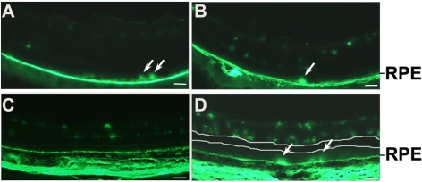
Purpose: The outer blood-retina barrier (BRB) separates the neural retina from the choroidal vasculature, which is responsible for approximately 80% of blood supplies in the eye. To determine the significance of outer BRB breakdown in diabetic retinopathy, the outer BRB-specific leakage of macromolecules in diabetic and ischemic rodents was investigated.
Methods: Diabetes and ischemia were induced in rodents by streptozotocin and oxygen-induced retinopathy, respectively. Diabetic and ischemic rodents were injected intravenously with fluorescein isothiocyanate (FITC)-dextran. The outer BRB-specific leakage in diabetic and ischemic rodents was visualized by fluorescent microscopy.
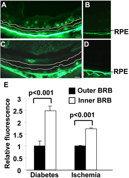
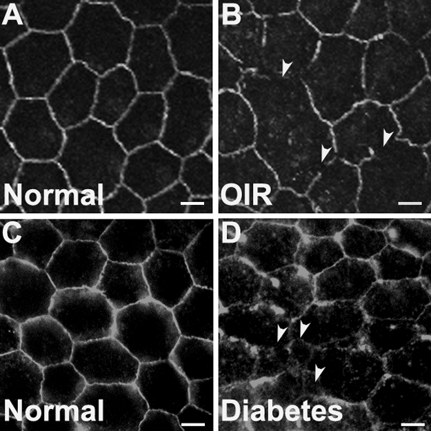
Results: A microscopic imaging assay was developed to examine outer BRB breakdown. The outer BRB-specific leakage of fluorescent macromolecules was visualized in diabetic and ischemic rodents. Substantial leakages of macromolecules through the outer BRB in diabetic and ischemic rodents were detected with this assay. The number of severe outer BRB leakage sites is inversely proportional to the size of macromolecules. Significant depletion of occludin in the RPE of ischemic and diabetic rodents was also observed.
Conclusions: For the first time, a microscopic imaging assay for directly visualizing macromolecules leaked through the outer BRB in rodents was developed. Using this assay, the authors demonstrated the significance of outer BRB breakdown in diabetes and ischemia, which will have implications to the understanding, diagnosis, and treatment of diabetic macular edema and other ocular diseases with outer BRB defects. The microscopic imaging assay established in this study will likely be very useful to the development of drugs for macular edema.
Figures

References
-
- Gordon WC, Rodriguez de Turco EB, Bazan NG. Retinal pigment epithelial cells play a central role in the conservation of docosahexaenoic acid by photoreceptor cells after shedding and phagocytosis. Curr Eye Res. 1992;11:73–83 - PubMed
-
- Benolken RM, Anderson RE, Wheeler TG. Membrane fatty acids associated with the electrical response in visual excitation. Science. 1973;182:1253–1254 - PubMed
-
- Philp NJ, Ochrietor JD, Rudoy C, Muramatsu T, Linser PJ. Loss of MCT1, MCT3, and MCT4 expression in the retinal pigment epithelium and neural retina of the 5A11/basigin-null mouse. Invest Ophthalmol Vis Sci. 2003;44:1305–1311 - PubMed
Publication types
MeSH terms
Substances
Grants and funding
LinkOut - more resources
Full Text Sources
Other Literature Sources
Medical
