. 2011 Jul;4(4):397-408.
doi: 10.1038/mi.2010.82. Epub 2010 Dec 22.

Homeostatic control of conjunctival mucosal goblet cells by NKT-derived IL-13

Affiliations

Homeostatic control of conjunctival mucosal goblet cells by NKT-derived IL-13

C S De Paiva et al. Mucosal Immunol. 2011 Jul.

Abstract

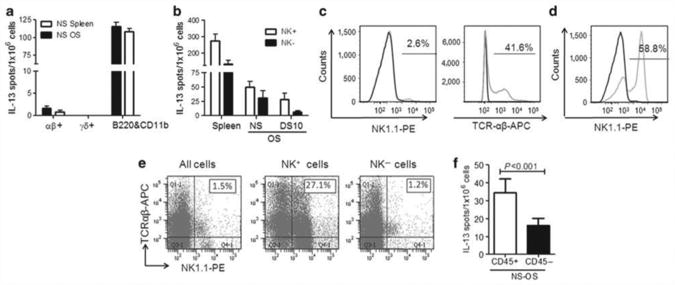
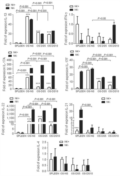
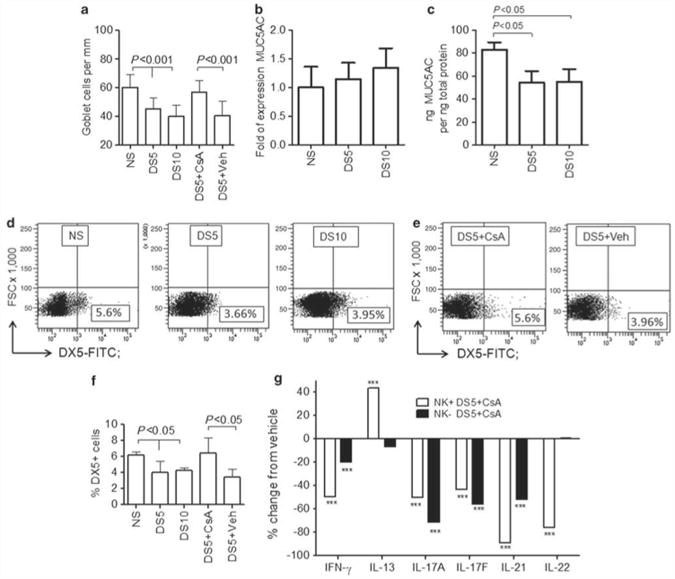
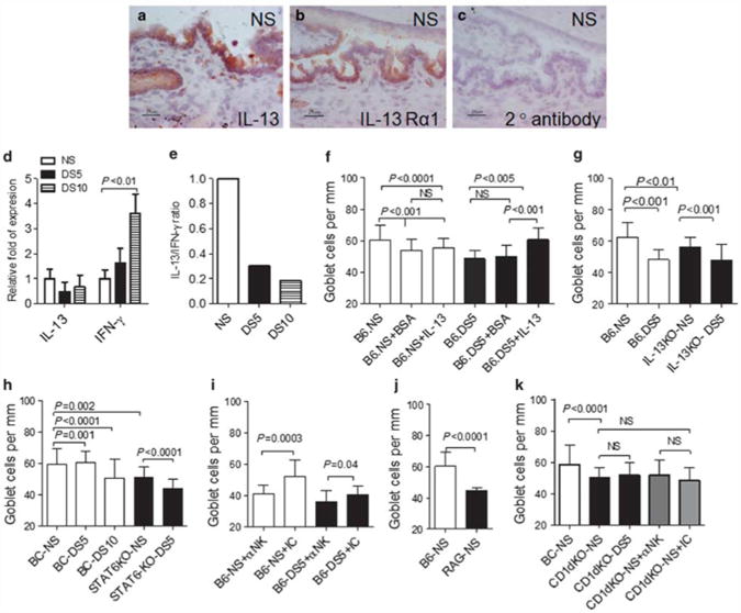
Although the effects of the interleukin 13 (IL-13) on goblet cell (GC) hyperplasia have been studied in the gut and respiratory tracts, its effect on regulating conjunctival GC has not been explored. The purpose of this study was to determine the major IL-13-producing cell type and the role of IL-13 in GC homeostasis in normal murine conjunctiva. Using isolating techniques, we identified natural killer (NK)/natural killer T (NKT) cells as the main producers of IL-13. We also observed that IL-13 knockout (KO) and signal transducer and activator of transcription 6 knockout (STAT6KO) mice had a lower number of periodic acid Schiff (PAS)+GCs. We observed that desiccating stress (DS) decreases NK population, GCs, and IL-13, whereas it increases interferon-γ (IFN-γ) mRNA in conjunctiva. Cyclosporine A treatment during DS maintained the number of NK/NKT cells in the conjunctiva, increased IL-13 mRNA in NK+ cells, and decreased IFN-γ and IL-17A mRNA transcripts in NK+ and NK- populations. C57BL/6 mice chronically depleted of NK/NKT cells, as well as NKT cell-deficient RAG1KO and CD1dKO mice, had fewer filled GCs than their wild-type counterparts. NK depletion in CD1dKO mice had no further effect on the number of PAS+ cells. Taken together, these findings indicate that NKT cells are major sources of IL-13 in the conjunctival mucosa that regulates GC homeostasis.


Conflict of interest statement

Disclosure: The authors declared no conflict of interest.

Figures

Figure 1
Figure 1
NK and NKT cells produce IL-13. (a) Mean±s.d. of interleukin 13 (IL-13) enzyme-linked immunosorbent spots (ELISPOTs) after γδ + isolation from nonstressed (NS) ocular surface (OS) and NS spleen, showing the three isolated populations (αβ +, γδ +, and B220 and CD11b + cells). (b) Mean±s. d. of IL-13 ELISPOTs after natural killer (NK) isolation from NS and desiccated-stressed (DS) OS for 10 days (DS10) and NS spleen, showing the two isolated populations, NK positive ( + ) and NK negative ( − ). (c) Histogram of flow cytometry analysis of total splenocytes stained with NK1.1-phycoerythrin (PE)-conjugated antibody (gray line) or T-cell receptor αβ-antigen-presenting cell (TCRαβ-APC) and isotype control antibody (black line). The number over line indicates percentage of positive cells over the isotype control antibody. (d, e) Flow cytometry analysis. Lymphocytes were gated based on characteristic light-scatter properties, and single lymphocytes were gated based on forward scatter height vs. forward scatter area (FSC-A). The numbers in the quadrants or over line indicate the percentage of cells. (d) Histogram of flow cytometry analysis of NK-positive cells stained with NK1.1-PE-conjugated antibody (gray line) or isotype control antibody (black line). The number over line indicates percentage of positive cells over the isotype control antibody. (e) Flow cytometry analysis of freshly isolated NK cells dual stained with NK1.1-PE + TCRαβ in all populations (all cells=no fraction, NK +, and NK − cells) from NS spleens. Percentage of double-positive cells is indicated in top right quadrant. (f) Mean±s.d. of IL-13 ELISPOTs after CD45 isolation from NS OS.
Figure 2
Figure 2
Gene expression analysis of isolated NK populations. Relative fold expression in natural killer (NK)/natural killer T (NKT)-positive and -negative populations. mRNA transcript levels of interleukin 13 (IL-13), interferon-γ (IFN-γ), IL-17A, IL-17F, IL-22, IL-21, and IL-4 in freshly isolated NK positive (NK +) and negative (NK −) cells isolated cells from nonstressed (NS) and desiccating-stressed (DS) ocular surface (OS) for 5 (DS5) or 10 days (DS10) and normal spleen. *** P< 0.001, comparison of NK + vs. NK − populations within the tissue. Comparison of different NK populations across different time points is shown by numerical values in the graph.
Figure 3
Figure 3
Effects of CsA treatment on NK and NKT cells. (a) Mean±s.d. of conjunctival goblet cell (GC) density in nonstressed (NS) C57BL/6 control mice and mice subjected to desiccating stress for 5 or 10 days (DS5 and DS10, respectively) or DS5 treated with Cyclosporine A (DS5 + CsA) or DS5 treated with CsA vehicle (DS5 + Veh). (b) mRNA transcript levels of MUC5AC in conjunctiva of NS C57BL/6 control mice and mice subjected to DS5 and DS10, respectively. (c) Mean±.d. of protein concentrations of MUC5AC in conjunctiva of NS C57BL/6 control mice and mice subjected to DS5 and DS10, respectively. (d, e) Representative flow cytometry analysis of freshly isolated ocular surface cells stained with pan-NK marker DX5 –fluorescein isothiocyanate (FITC)-conjugated antibody in NS C57BL/6 controls mice and mice subjected to DS5 and DS10, respectively (d) or DS5 treated with CsA (DS5 + CsA) or DS5 treated with CsA vehicle (DS5 + Veh) (e). Lymphocytes were gated based on characteristic light-scatter properties, and single lymphocytes were gated based on forward scatter height vs. forward scatter area (FSC-A). Numbers in the quadrants indicate the percentage of cells. (f) Mean±s.d. of the percentage of DX5+ cells evaluated by flow cytometry in three independent experiments. (g) Percentage change in the level of expression of interferon-γ (IFN-γ), IL-13, IL-17A, IL-21, and IL-22 mRNA transcripts in CsA-treated NK + and NK − cell populations, compared with vehicle. *** P< 0.001, comparison of NK + or NK − DS5 + CSA vs. the same population from DS5 + vehicle.
Figure 4
Figure 4
IL-13 expression and goblet cell analysis. (a–c) Immunohistochemical staining for interleukin 13 (IL-13; red, in a) and IL-13 receptor α1 (IL-13Rα1; red, in b) and goat anti-rabbit secondary antibody alone (c) in conjunctiva sections of nonstressed (NS) control mice. Scale bar = 25 μm. (d) mRNA transcript levels of IL-13 and interferon-γ (IFN-γ) in conjunctiva in NS control mice and mice subjected to desiccating stress for 5 or 10 days (DS5 and DS10, respectively). (e) Ratio of IL-13/IFN-γ mRNA transcripts in conjunctiva. (f) Mean±s.d. of goblet cell (GC) density in NS C57BL/6 (B6) control mice and mice subjected to DS5 and mice after subconjunctival injection of BSA (+ BSA) or IL-13 (+ IL13). (g) Mean±s.d. of GC density in NS C57BL/6 (B6) controls mice and mice subjected to DS5 and IL-13 knockout (KO) mice. (h) Mean±s.d. of GC density in NS Bal/c (BC) control mice and mice subjected to DS5 and DS10 and signal transducer and activator of transcription 6 knockout (STAT6KO) at baseline and after DS5. (i) Mean±s. d. of GC density in NS C57BL/6 mice (B6) that received systemic injection of neutralizing antibody (NK1.1) to natural killer (NK) cells (αNK) or isotype control (IC) antibody before NS and after DS5. (j) Mean±s.d. of GC density in NS C57BL/6 (B6) control and RAG1KO mice. (k) Mean±s.d. of GC density in NS Bal/c (BC) control mice and CD1dKO at baseline, NS, and after DS5. A separate group NS CD1dKO mice received systemic injection of depleting antibody (NK1.1) to NK cells (αNK) or IC antibody.

References

    1. Nakamura T, Nishida K, Dota A, Matsuki M, Yamanishi K, Kinoshita S. Elevated expression of transglutaminase 1 and keratinization-related proteins in conjunctiva in severe ocular surface disease. Invest Ophthalmol Vis Sci. 2001;42:549–556. - PubMed
    1. Pflugfelder SC, Tseng SCG, Yoshino K, Monroy D, Felix C, Reis BL. Correlation of goblet cell density and mucosal epithelial membrane mucin expression with rose bengal staining in patients with ocular irritation. Ophthalmology. 1997;104:223–235. - PubMed
    1. Shahzeidi S, Aujla PK, Nickola TJ, Chen Y, Alimam MZ, Rose MC. Temporal analysis of goblet cells and mucin gene expression in murine models of allergic asthma. Exp Lung Res. 2003;29:549–565. - PubMed
    1. Shim JJ, et al. IL-13 induces mucin production by stimulating epidermal growth factor receptors and by activating neutrophils. Am J Physiol Lung Cell Mol Physiol. 2001;280:L134–L140. - PubMed
    1. Kondo M, et al. Elimination of IL-13 reverses established goblet cell metaplasia into ciliated epithelia in airway epithelial cell culture. Allergol Int. 2006;55:329–336. - PubMed

Publication types

MeSH terms