

Review
doi: 10.1038/eye.2010.149.
Epub 2010 Oct 29.
Retinal light toxicity
Affiliations
- PMID: 21178995
- PMCID: PMC3144654
- DOI: 10.1038/eye.2010.149
Item in Clipboard
Review
Retinal light toxicity
Eye (Lond).
2011 Jan.
Abstract
The ability of light to enact damage on the neurosensory retina and underlying structures has been well understood for hundreds of years. While the eye has adapted several mechanisms to protect itself from such damage, certain exposures to light can still result in temporal or permanent damage. Both clinical observations and laboratory studies have enabled us to understand the various ways by which the eye can protect itself from such damage. Light or electromagnetic radiation can result in damage through photothermal, photomechanical, and photochemical mechanisms. The following review seeks to describe these various processes of injury and many of the variables, which can mitigate these modes of injury.
Figures
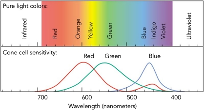
The portion of the electromagnetic spectrum that interacts with the eye is referred to as optical radiation and includes wavelengths from ultraviolet (100–400 nm), visible (400–760 nm), and infrared light (760–10 000+ nm). How Things Work: The Physics of Everyday Life, 3rd edn; Louis A Bloomfield; Copyright Wiley 2005. Reprinted with permission of John Wiley & Sons, Inc.
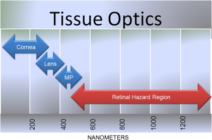
Schematic representation of the tissue optics of the human eye. The cornea, lens, and macular pigment (MP) absorb electromagnetic radiation preventing potential photic energy from high-energy, short-wavelength light. The retinal hazard region represents electromagnetic radiation not absorbed by the aforementioned ocular tissue.
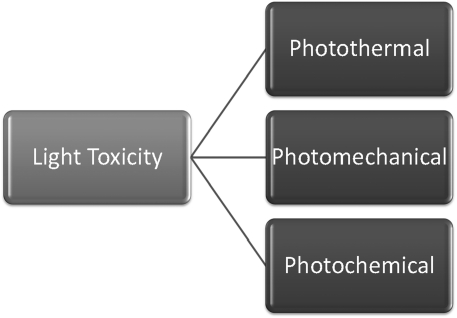
Schematic representation of the three major forms of photic injury.
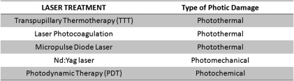
The ability of light to cause photic damage to the retina is utilized in several different types of laser treatments. Through either photothermal, photomechanical, or photochemical mechanisms, laser can be used to treat various ocular pathology.
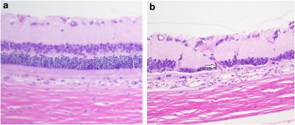
Normal histology of albino rat retina (a). Histopathology of abnormal rat retina exhibiting the development of atrophy and choroidal neovascularization (arrow) after several months of intense cyclic light exposure (b). Courtesy of Richard R Dubielzig, DVM, School of Veterinary Medicine, University of Wisconsin.
References
-
- Favazza AR. Literature on sun gazing. Am J Psychiatry. 1991;148 (2:281–282. - PubMed
-
- Noell WK, Walker VS, Kang BS, Berman S. Retinal damage by light in rats. Invest Ophthalmol. 1966;5 (5:450–473. - PubMed
-
- Green WR, Robertson DM. Pathologic findings of photic retinopathy in the human eye. Am J Ophthalmol. 1991;112 (5:520–527. - PubMed
Publication types
MeSH terms
LinkOut - more resources
Full Text Sources
Other Literature Sources
Medical
