

Review
doi: 10.1038/nrm3028.
Mechanisms of mitophagy
Affiliations
- PMID: 21179058
- PMCID: PMC4780047
- DOI: 10.1038/nrm3028
Item in Clipboard
Review
Mechanisms of mitophagy
Nat Rev Mol Cell Biol.
2011 Jan.
Abstract
Autophagy not only recycles intracellular components to compensate for nutrient deprivation but also selectively eliminates organelles to regulate their number and maintain quality control. Mitophagy, the specific autophagic elimination of mitochondria, has been identified in yeast, mediated by autophagy-related 32 (Atg32), and in mammals during red blood cell differentiation, mediated by NIP3-like protein X (NIX; also known as BNIP3L). Moreover, mitophagy is regulated in many metazoan cell types by parkin and PTEN-induced putative kinase protein 1 (PINK1), and mutations in the genes encoding these proteins have been linked to forms of Parkinson's disease.
Figures
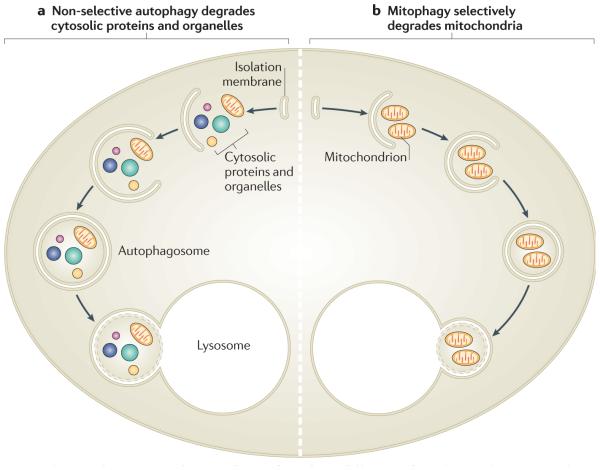
a | Non-selective autophagy occurs when cells are deprived of nutrients. It degrades a range of cytosolic contents, including proteins and many types of organelles. After their recruitment into isolation membranes, cytosolic components are sealed into autophagosomes that fuse with lysosomes. The degradation of these components in the lysosome supplies building blocks for re-use and for metabolism to provide ATP. b | By contrast, mitophagy occurs to eliminate mitochondria, either to regulate their number or to specifically remove ones that are damaged. Mitochondria are selectively recruited into isolation membranes, which seal and then fuse with lysosomes to eliminate the trapped mitochondria.
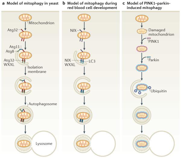
Mitophagy requires the specific labelling of mitochondria and their recruitment into isolation membranes. a | In yeast this occurs through the outer mitochondrial membrane protein autophagy-related 32 (Atg32), which can bind the isolation membrane protein Atg8 through its WXXL-like motif. How mitochondria are specified for removal to calibrate the mitochondrial population density according to the cell’s metabolic demand remains unknown but may occur through the adaptor protein Atg11, which binds both Atg32 and Atg8 and could physically link mitochondria and isolation membranes. Finally, the isolation membranes seal mitochondria in autophagosomes and fuse with lysosomes to allow degradation. b | During differentiation, red blood cells lose their mitochondria through mitophagy. Expression of the outer mitochondrial membrane protein NIP3-like protein X (NIX; also known as BNIP3L) increases during red blood cell differentiation and is required for mitochondrial removal. NIX has a WXXL-like motif, which binds to MAP1 light chain 3 (LC3; a homologue of yeast Atg8) on isolation membranes and is thought to mediate the binding and sequestration of mitochondria into autophagosomes. c | When mitochondria are damaged and lose membrane potential, the kinase PTEN-induced putative kinase protein 1 (PINK1) accumulates (shown as a halo around the mitochondrion) and recruits the E3 ubiquitin ligase parkin from the cytosol specifically to the damaged mitochondrion. Parkin ubiquitylates mitochondrial proteins and causes mitochondria to become engulfed by isolation membranes that then fuse with lysosomes. This may mediate mitochondrial quality control.
References
-
- Nakatogawa H, Suzuki K, Kamada Y, Ohsumi Y. Dynamics and diversity in autophagy mechanisms: lessons from yeast. Nature Rev. Mol. Cell Biol. 2009;10:458–467. - PubMed
Publication types
MeSH terms
Grants and funding
LinkOut - more resources
Full Text Sources
Other Literature Sources
Molecular Biology Databases
Research Materials
