. 2010 Dec 23;468(7327):1115-8.
doi: 10.1038/nature09599.

S-glutathionylation uncouples eNOS and regulates its cellular and vascular function

Affiliations

S-glutathionylation uncouples eNOS and regulates its cellular and vascular function

Chun-An Chen et al. Nature. .

Abstract

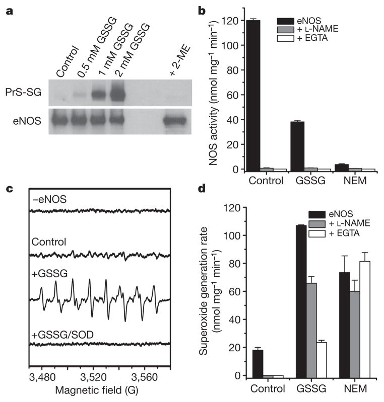
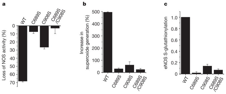
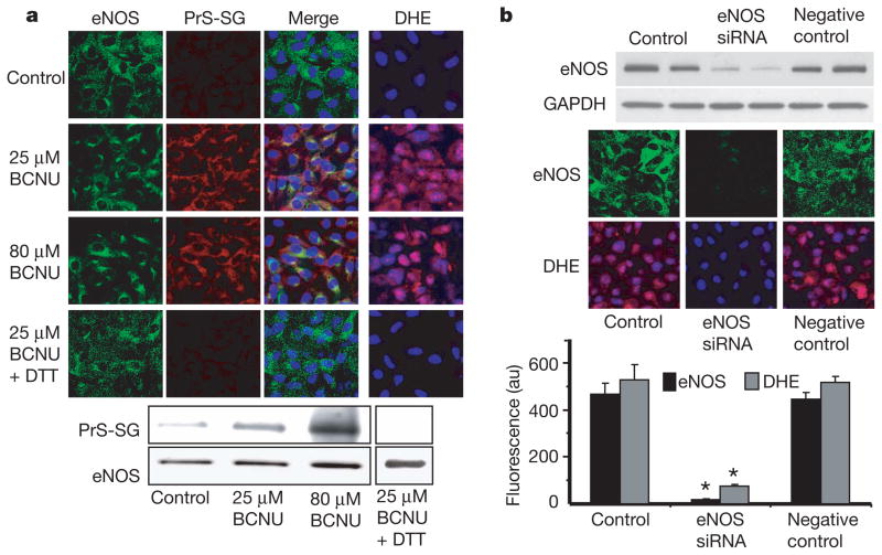
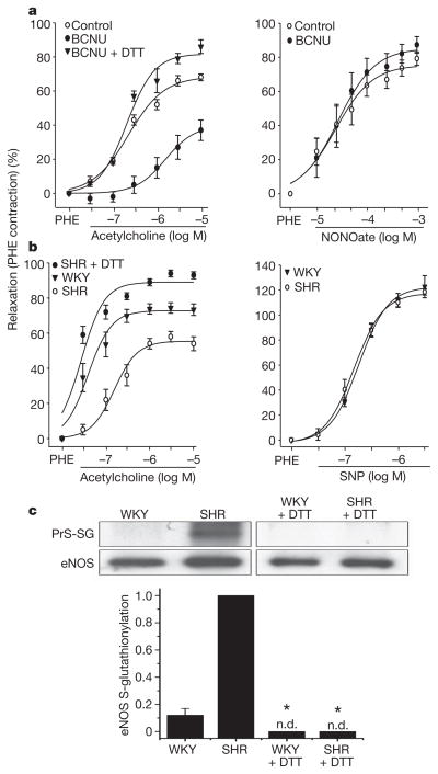
Endothelial nitric oxide synthase (eNOS) is critical in the regulation of vascular function, and can generate both nitric oxide (NO) and superoxide (O(2)(•-)), which are key mediators of cellular signalling. In the presence of Ca(2+)/calmodulin, eNOS produces NO, endothelial-derived relaxing factor, from l-arginine (l-Arg) by means of electron transfer from NADPH through a flavin containing reductase domain to oxygen bound at the haem of an oxygenase domain, which also contains binding sites for tetrahydrobiopterin (BH(4)) and l-Arg. In the absence of BH(4), NO synthesis is abrogated and instead O(2)(•-) is generated. While NOS dysfunction occurs in diseases with redox stress, BH(4) repletion only partly restores NOS activity and NOS-dependent vasodilation. This suggests that there is an as yet unidentified redox-regulated mechanism controlling NOS function. Protein thiols can undergo S-glutathionylation, a reversible protein modification involved in cellular signalling and adaptation. Under oxidative stress, S-glutathionylation occurs through thiol-disulphide exchange with oxidized glutathione or reaction of oxidant-induced protein thiyl radicals with reduced glutathione. Cysteine residues are critical for the maintenance of eNOS function; we therefore speculated that oxidative stress could alter eNOS activity through S-glutathionylation. Here we show that S-glutathionylation of eNOS reversibly decreases NOS activity with an increase in O(2)(•-) generation primarily from the reductase, in which two highly conserved cysteine residues are identified as sites of S-glutathionylation and found to be critical for redox-regulation of eNOS function. We show that eNOS S-glutathionylation in endothelial cells, with loss of NO and gain of O(2)(•-) generation, is associated with impaired endothelium-dependent vasodilation. In hypertensive vessels, eNOS S-glutathionylation is increased with impaired endothelium-dependent vasodilation that is restored by thiol-specific reducing agents, which reverse this S-glutathionylation. Thus, S-glutathionylation of eNOS is a pivotal switch providing redox regulation of cellular signalling, endothelial function and vascular tone.


Figures

Figure 1
Figure 1. S-glutathionylation of heNOS occurs and inhibits NOS activity
a, Immunoblotting of heNOS S-glutathionylation. Top: immunoblotting of protein S-glutathionylation (PrS-SG) with anti-GSH antibody. Control non-S-glutathionylated heNOS (1 μg in 20 μl) or heNOS S-glutathionylated by 0.5, 1 or 2 mM GSSG at room temperature (23 °C) for 1 h. Treatment with 2-mercaptoethanol (ME) after S-glutathionylation with 2 mM GSSG reversed the S-glutathionylation. Bottom: immunoblotting with anti-eNOS antibody. b, Effect of S-glutathionylation and S-alkylation on heNOS activity. NOS activity was measured from control, S-glutathionylated (2 mM GSSG for 20 min) or alkylated (1 mM NEM for 20 min) heNOS. NOS activity of treated or untreated heNOS was fully inhibited by L-NAME (1 mM) or EGTA (1 mM). c, d, Effects of S-glutathionylation on O2•− generation from heNOS. O2•− generation was measured from control, S-glutathionylated (as in b) or alkylated (as in b) BH4-bound heNOS by EPR spin trapping with 25 mM 5-diethoxyphosphoryl-5-methyl-1-pyrroline N-oxide (DEPMPO). c, Spin-trapping showed no signal in the absence of heNOS and only trace signal from control enzyme; S-glutathionylation triggered a marked increase in O2•− generation with a O2•−-adduct spectrum that was quenched by Cu, Zn superoxide dismutase (SOD) (200 U ml−1). d, Effect of L-NAME (1 mM) and EGTA (1 mM) on O2•− generation from control, S-glutathionylated and alkylated BH4-bound heNOS. Results in b and d are shown as means and s.e.m. (n = 3–5).
Figure 2
Figure 2. Cysteine mutants (C689S, C908S and C689S/C908S) of heNOS resist S-glutathionylation and secondary uncoupling
WT heNOS and heNOS C689S, C908S and C689S/C908S mutants were treated with 2 mM GSSG. a, Percentage loss of NOS activity after treatment of heNOS with GSSG. b, Percentage increase in O2•− generation after treatment of heNOS with GSSG. c, Ratio of relative eNOS S-glutathionylation to eNOS protein. The relative intensity of eNOS S-glutathionylation/eNOS protein was normalized to the wild-type value. The Cys→Ser mutants maintained a NOS activity similar to that of the wild type (120 ± 12 n mol min−1 mg−1). Results are shown as means and s.e.m. (n =3).
Figure 3
Figure 3. Effect of redox stress on eNOS S-glutathionylation and function in endothelial cells
a, Immunostaining of eNOS (left column, green fluorescence) and S-glutathionylation (second column, red fluorescence) in control BAECs and cells preincubated with BCNU. The third column shows the merged S-glutathionylation/eNOS image along with 4′,6-diamidino-2-phenylindole (DAPI) staining of the nucleus (blue). eNOS staining and S-glutathionylation seem to co-localize. The right-hand column shows O2•− detection with dihydroethidine (DHE), which is oxidized by O2•− to a product with red fluorescence, and the cell nuclei were counterstained with DAPI (blue). Increased O2•− generation was seen in the BCNU-treated cells. Bottom: immunoprecipitation of eNOS from the BCNU-treated cells shows that eNOS S-glutathionylation occurs but is reversed by 1 mM DTT. The upper row shows immunoblotting with anti-GSH antibody; the lower row shows immunoblotting with anti-eNOS antibody. b, Effects of eNOS silencing from BAECs on BCNU-induced O2•− generation. Top: immunoblotting against eNOS to determine the efficiency of eNOS silencing. Middle: confocal microscopy of eNOS (upper row) and O2•− measurements with DHE (lower row). NOS3 short interfering RNA (siRNA) greatly decreases eNOS expression in BAECs (middle column). Bottom: graph of the effect of eNOS silencing on BCNU-induced O2•− generation. Results are shown as means and s.e.m. (n =5). Asterisk, P < 0.001.
Figure 4
Figure 4. Effect of redox stress on eNOS S-glutathionylation and function in vessels
a, Endothelium-dependent and endothelium-independent vasorelaxation in control and BCNU (80 μM)-treated rat aortic rings. BCNU markedly decreased endothelium-dependent relaxation to acetylcholine (left panel) but not endothelium-independent relaxation by the NO donor NONOate (right panel). DTT (1 mM for 20 min) reversed the BCNU-induced inhibition of relaxation (left panel). Aortic relaxation is plotted as the percentage decrease in phenylephrine (PHE)-induced contraction against agonist concentration on a logarithmic scale. Results are shown as means ± s.e.m.; P < 0.05, BCNU versus control or BCNU + DTT (n =4). b, Endothelium-dependent vasorelaxation in spontaneously hypertensive (SHR) and control (WKY) aortic rings. SHR rings showed a marked decrease in relaxation to acetylcholine; however, DTT (as above) re-established the acetylcholine response. Endothelium-independent relaxation (right) was similar for both SHR and WKY rings. Aortic relaxation is expressed as in a. P < 0.05, SHR versus WKY or SHR + DTT (n =4). See also Supplementary Fig. 10. c, eNOS S-glutathionylation of SHR and WKY aortae. Top: WKY and SHR aortae, either untreated or DTT-pretreated as in b, were homogenized. This was followed by immunoprecipitation with anti-eNOS antibody. The immunoprecipitation products were separated by SDS–PAGE followed by immunoblotting against anti-GSH and anti-eNOS antibodies. In SHR aortae, eNOS S-glutathionylation was markedly increased compared with WKY aortae and was abolished by pretreatment with DTT. Bottom: ratio of relative intensity of eNOS S-glutathionylation/eNOS, normalized to SHR aortae. There is only trace eNOS S-glutathionylation in WKY aortae, whereas high levels are seen in SHR aortae. There was no detectable (n.d.) NOS S-glutathionylation in DTT-pretreated WKY or SHR aortae. Asterisk, P < 0.001 versus SHR (n =5).

References

    1. Bredt DS, et al. Cloned and expressed nitric oxide synthase structurally resembles cytochrome P-450 reductase. Nature. 1991;351:714–718. - PubMed
    1. Palmer RM, Ashton DS, Moncada S. Vascular endothelial cells synthesize nitric oxide from L-arginine. Nature. 1988;333:664–666. - PubMed
    1. Rapoport RM, Draznin MB, Murad F. Endothelium-dependent relaxation in rat aorta may be mediated through cyclic GMP-dependent protein phosphorylation. Nature. 1983;306:174–176. - PubMed
    1. Xia Y, Tsai AL, Berka V, Zweier JL. Superoxide generation from endothelial nitric-oxide synthase. A Ca2+/calmodulin-dependent and tetrahydrobiopterin regulatory process. J Biol Chem. 1998;273:25804–25808. - PubMed
    1. Vasquez-Vivar J, et al. Superoxide generation by endothelial nitric oxide synthase: the influence of cofactors. Proc Natl Acad Sci USA. 1998;95:9220–9225. - PMC - PubMed

Publication types

MeSH terms