. 2010 Dec 14;5(12):e14320.
doi: 10.1371/journal.pone.0014320.

Iota-carrageenan is a potent inhibitor of influenza A virus infection

Affiliations

Iota-carrageenan is a potent inhibitor of influenza A virus infection

Andreas Leibbrandt et al. PLoS One. .

Abstract

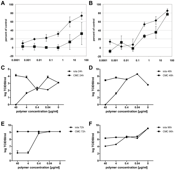
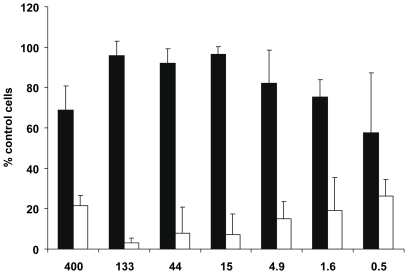
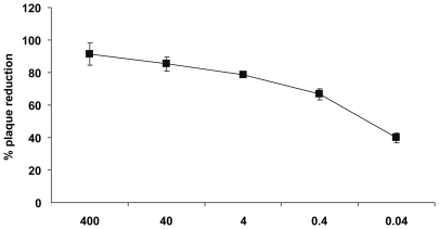
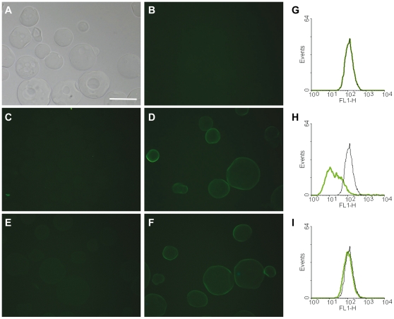
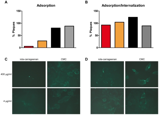
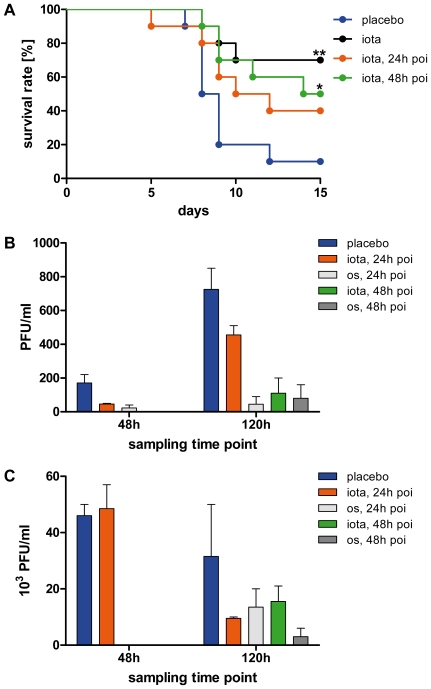
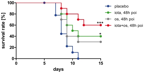
The 2009 flu pandemic and the appearance of oseltamivir-resistant H1N1 influenza strains highlight the need for treatment alternatives. One such option is the creation of a protective physical barrier in the nasal cavity. In vitro tests demonstrated that iota-carrageenan is a potent inhibitor of influenza A virus infection, most importantly also of pandemic H1N1/2009 in vitro. Consequently, we tested a commercially available nasal spray containing iota-carrageenan in an influenza A mouse infection model. Treatment of mice infected with a lethal dose of influenza A PR8/34 H1N1 virus with iota-carrageenan starting up to 48 hours post infection resulted in a strong protection of mice similar to mice treated with oseltamivir. Since alternative treatment options for influenza are rare, we conclude that the nasal spray containing iota-carrageenan is an alternative to neuraminidase inhibitors and should be tested for prevention and treatment of influenza A in clinical trials in humans.


Conflict of interest statement

Competing Interests: The authors AL, AG, CM, MK, RW, BP, BF, PG, and EP are employed by Marinomed. Authors HU, EP and AG are co-founders of Marinomed. AG and EP are inventors of patent # WO2009027057 held by Marinomed Biotechnologie GmbH that relates to the content of the manuscript. Marinomed Biotechnologie GmbH is financing the processing charge of this manuscript. TF has previously served as principal investigator for a clinical investigator-initiated study that was co-financed by Marinomed. This does not alter the authors' adherence to all the PLoS ONE policies on sharing data and materials. Authors MB and DK declare no competing interests.

Figures

Figure 1
Figure 1. Iota-carrageenan promotes cell viability and reduces viral titer of influenza A-infected MDCK cells.
MDCK cells grown in 96-well plates were infected with H1N1 A/PR/8/34 virus (A) and H3N2 A/Aichi/2/68 virus (B) (0.01 PFU/cell) in the presence of carrageenans (iota-carrageenan black diamonds, kappa-carrageenan black squares) at concentrations as indicated on the x-axis in µg/ml. Plates were incubated at 37°C until cells in the control (no polymer added) showed >90% damage. Cell proliferation was determined with a Resazurin-based in vitro toxicology assay. Samples were measured fluorometrically by monitoring the increase in fluorescence at a wavelength of 590 nm using an excitation wavelength of 544 nm. Values obtained from mock-infected cells were set to 100%, and the values of cells infected in the absence of polymer were set to 0% (y-axis). (C)-(F) MDCK cells were infected with H1N1 A/PR/8/34 as before and further kept in the presence of iota-carrageenan (circles) or the control polymer CMC (squares) at indicated concentrations and 24 (C), 48 (D), 72 (E), and 96 hours (F) post infection, respectively. Supernatants were harvested, pooled, and subsequently used to determine the TCID50/ml according to the method of Reed and Muench . The points represent the mean of a quadruplicate experiment, the standard deviation is indicated.
Figure 2
Figure 2. Effect of iota-carrageenan on influenza A-infected primary human nasal epithelial cells.
Primary human nasal epithelial cells (HNep) cells grown in 96-well plates were infected with A/PR/8/34 virus (5 PFU/cell) in the presence of iota-carrageenan at concentrations indicated on the x-axis in µg/ml. 30 minutes after infection, the inoculum was removed and medium containing iota-carrageenan (black bars) or CMC (white bars) in indicated concentrations added. Cell proliferation was determined with a Resazurin-based in vitro toxicology assay. Samples were measured fluorometrically by monitoring the increase in fluorescence at a wavelength of 590 nm using an excitation wavelength of 544 nm. Values obtained from mock-infected cells were set to 100%, and the values of cells infected in the absence of polymer were set to 0% (y-axis). The bars represent the mean of a quadruplicate experiment, the standard deviation is indicated.
Figure 3
Figure 3. Effect of iota-carrageenan on pandemic H1N1/2009 virus.
Confluent monolayers of MDCK cells in 6-well plates were washed free of protein-containing growth medium before use. An equal volume of virus suspension mixed with iota-carrageenan, containing 50 to 150 plaque-forming units, was added 5 to 10 min later, and plates were incubated at room temperature for 60 min with frequent shaking. The inoculum was removed and covered with an overlay medium consisting of 0.6% agarose (3 ml) in Eagle minimal essential medium and trypsin (2 µg/ml). Plates were incubated at 37°C in a humidified atmosphere with 5% CO2. After 36 to 48 h, plaques were stained with crystal violet and counted. The percentage of plaque inhibition relative to infected control plates (y-axis) was determined for each drug concentration (x-axis). The standard deviation of three independent experiments is indicated.
Figure 4
Figure 4. Binding of H1N1 influenza virus to iota-carrageenan.
(A)-(F). Alexa Fluor 488-conjugated H1N1 influenza virus (H1N1-A488) was incubated with iota-carrageenan-coated agarose beads (iota-beads) or control beads for 30 min at room temperature and visualized microscopically. (A) Bright field picture of iota-beads, showing no green auto-fluorescence (B). (C) Control agarose beads incubated with H1N1-A488 do not facilitate unspecific virus binding. (D) Iota-beads incubated with H1N1-A488 demonstrates binding of virus to iota-carrageenan as evidenced by bright green staining of iota-beads. (E) Binding of H1N1-A488 to iota-beads is inhibited in the presence of iota-carrageenan (400 µg/ml), but is not abolished in the presence of CMC (400 µg/ml) (F). Scale bar  = 100 µm. (G) FACS analysis of MDCK cells incubated with H1N1-A488 in the presence of iota-carrageenan (400 µg/ml) (H) or control polymer CMC (400 µg/ml) (I) showing that binding of H1N1-A488 to cognate receptors is inhibited by iota-carrageenan but not CMC.
Figure 5
Figure 5. Effect of iota-carrageenan on H1N1 virus adsorption and internalization.
(A) Adsorption. H1N1 virus was added to MDCK cells in the presence of different concentrations of iota-carrageenan or control polymer carboxymethylcellulose (CMC). After viral adsorption for 1 h at 4°C, cells were washed and the number of cell-bound infectious viral particles determined by plaque assay; red bar 400 µg/ml, orange bar 4 µg/ml iota-carrageenan, black bar 400 µg/ml, grey bar 4 µg/ml CMC. (B) Adsorption/Internalization. H1N1 virus was added to MDCK cells and adsorbed for 1 h at 4°C. Cells were washed and allowed to internalize virus in the presence or absence of different concentrations of iota-carrageenan or CMC for 1 h at 37°C. Subsequently, internalized infectious viral particles were determined by plaque assay. (C) Immunofluorescent visualisation of virus adsorption in the presence of iota-carrageenan or CMC. 1 h post adsorption at 4°C, cells were stained after 1 h at 37°C with a mouse anti-NP antibody. (D) Adsorption/Internalization. H1N1 was added to MDCK cells and adsorbed for 1 h at 4°C. Cells were washed and allowed to internalize virus in the presence of iota-carrageenan or CMC for 1 h at 37°C. Compare the bright green stainings in Figure 5D indicative of productive infection to 5C, where no green fluorescence is detected at high iota-carrageenan concentration.
Figure 6
Figure 6. Therapeutic efficacy of iota-carrageenan against H1N1 influenza virus in a lethal mouse infection model.
(A) Ten mice per group were intranasally infected with 8.7×10² PFU H1N1 A/PR/8/34 viral particles at day 0. Intranasal therapy twice daily with 60 µg iota-carrageenan in 0.5% NaCl or placebo (blue) started on the same day as infection (black), 24 h post infection (poi) (orange), or 48 h poi (green), and was performed twice daily for the entire experiment. P values were calculated by a Log-rank (Mantel-Cox) test. Asterisk, p<0.05, double asterisk p<0.01. (B)-(C). Determination of viral titers from nose (B) and lung (C) specimens. Five mice per group were intranasally infected at day 0 as before. The group receiving placebo (blue) was compared to groups receiving intranasal therapy with iota-carrageenan or oral therapy with oseltamivir (10 mg/kg/day in 5% sucrose) starting 24 (orange or light grey) and 48 hours (green or dark grey) post infection until groups of mice were sacrificed at day 2 and 5 days, respectively. Subsequently, nose and lung specimens of animals from each experimental group and time point were pooled and viral titers determined by plaque assays on MDCK cells at two different dilutions. Bars represent the mean±SEM.
Figure 7
Figure 7. Efficacy of iota-carrageenan in mice in comparison to oseltamivir.
Ten mice per group were intranasally infected with 8.7×10³ PFU H1N1/PR/8/34 viral particles at day 0 and therapy started 48 h poi (blue indicates the placebo treatment). In addition to the group with intranasal treatment twice daily with 60 µg iota-carrageenan (green), a group of mice also received an oral dose of oseltamivir (10 mg/kg/day in 5% sucrose) (grey) twice daily for 5 days, and accordingly in combination with iota-carrageenan (red). P values were calculated by a Log-rank (Mantel-Cox) test. Survival was monitored daily for 15 days. Asterisk, p<0.05; triple asterisk p<0.001.

References

    1. Hayden F. Developing new antiviral agents for influenza treatment: what does the future hold? Clin Infect Dis. 2009;48:3–13. - PubMed
    1. Pinto LH, Holsinger LJ, Lamb RA. Influenza virus M2 protein has ion channel activity. Cell. 1992;69:517–528. - PubMed
    1. Schnell JR, Chou JJ. Structure and mechanism of the M2 proton channel of influenza A virus. Nature. 2008;451:591–595. - PMC - PubMed
    1. Jefferson T, Demicheli V, Di Pietrantonj C, Rivetti D. Cochrane - Amantadine and rimantadine for influenza A in adults. Cochrane Database Syst Rev 2006 - PMC - PubMed
    1. Nelson MI, Simonsen L, Viboud C, Miller MA, Holmes EC. The origin and global emergence of adamantane resistant A/H3N2 influenza viruses. Virology. 2009;388:270–278. - PMC - PubMed

Publication types