. 2010 Dec 13;5(12):e15251.
doi: 10.1371/journal.pone.0015251.

The neurotoxicity of DOPAL: behavioral and stereological evidence for its role in Parkinson disease pathogenesis

Affiliations

The neurotoxicity of DOPAL: behavioral and stereological evidence for its role in Parkinson disease pathogenesis

W Michael Panneton et al. PLoS One. .

Erratum in

  • PLoS One. 2011;6(2). doi: 10.1371/annotation/819cbf4a-7e3e-4e4e-a449-b667bc6e8957

Abstract

Background: The etiology of Parkinson disease (PD) has yet to be fully elucidated. We examined the consequences of injections of 3,4-dihydroxyphenylacetaldehyde (DOPAL), a toxic metabolite of dopamine, into the substantia nigra of rats on motor behavior and neuronal survival.

Methods/principal findings: A total of 800 nl/rat of DOPAL (1 µg/200 nl) was injected stereotaxically into the substantia nigra over three sites while control animals received similar injections of phosphate buffered saline. Rotational behavior of these rats was analyzed, optical density of striatal tyrosine hydroxylase was calculated, and unbiased stereological counts of the substantia nigra were made. The rats showed significant rotational asymmetry ipsilateral to the lesion, supporting disruption of dopaminergic nigrostriatal projections. Such disruption was verified since the density of striatal tyrosine hydroxylase decreased significantly (p<0.001) on the side ipsilateral to the DOPAL injections when compared to the non-injected side. Stereological counts of neurons stained for Nissl in pars compacta of the substantia nigra significantly decreased (p<0.001) from control values, while counts of those in pars reticulata were unchanged after DOPAL injections. Counts of neurons immunostained for tyrosine hydroxylase also showed a significant (p=0.032) loss of dopaminergic neurons. In spite of significant loss of dopaminergic neurons, DOPAL injections did not induce significant glial reaction in the substantia nigra.

Conclusions: The present study provides the first in vivo quantification of substantia nigra pars compacta neuronal loss after injection of the endogenous toxin DOPAL. The results demonstrate that injections of DOPAL selectively kills SN DA neurons, suggests loss of striatal DA terminals, spares non-dopaminergic neurons of the pars reticulata, and triggers a behavioral phenotype (rotational asymmetry) consistent with other PD animal models. This study supports the "catecholaldehyde hypothesis" as an important link for the etiology of sporadic PD.


Conflict of interest statement

Competing Interests: The authors have declared that no competing interests exist.

Figures

Figure 1
Figure 1. Box plot illustrating the behavioral changes in rats after unilateral injections of DOPAL into their substantia nigra.
Rats showed rotational asymmetry, turning significantly towards the side of DOPAL injections. *p<0.05.
Figure 2
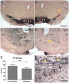
Figure 2. Photomicrographs of brain sections (case R2508) immunohistochemically-stained against tyrosine hydroxylase (TH) after injections of DOPAL into the substantia nigra, pars compacta (SNpc).
Note the gross reduction of TH immunoreactivity in the SN at the site of injection (B; yellow arrowhead) versus the non-injected side (A). Similar loss of TH staining is seen in the striatum ipsilateral to the injection (D, arrows) versus that on the non-injected side (C, arrows), suggesting disruption of nigral dopaminergic terminals. The area just lateral to the anterior commissure (D, yellow arrowhead) however was always densely labeled (see text). Densitometry of immunostaining of striatal TH (E) showed significant differences (p<0.001) of the whole striatum contralateral and ipsilateral to DOPAL injections. Spot density measurements of ventrolateral parts of the striatum (D, red circles), however, showed an 80% loss of immunoreactivity ipsilateral to the injection. Intensely stained neurons with antibodies against tyrosine hydroxylase (F, yellow arrowheads) were sometimes seen in the SNpc of control brains surrounded by numerous neurons stained only for Nissl (F, black arrows), suggesting that counting only TH-immunostained neurons may be problematic. Abbreviations: ac, anterior commissure; SNpc, pars compacta of substantia nigra; SNpr, pars reticulata of substantia nigra. *** p<0.001.
Figure 3
Figure 3. Photomicrographs of sections through the SN stained for Nissl with neutral red.
Red lines mark the boundaries enclosing the substantia nigra, pars compacta, while green lines encompass the substantia nigra, pars reticulata. Unbiased stereological counts using optical fractionator probes were made of neurons in both SNpc and SNpr in sections from animals injected with buffer (A; case R2546) and those injected with DOPAL (B, case R2505). Note the significant (p≤0.001) loss of SNpc neurons in rats (C) after the DOPAL injection when compared to control rats, while no loss of neurons was seen in the adjacent SNpr.
Figure 4
Figure 4. Brightfield photomicrographs of sections through rat brains stained immunohistochemically for glia.
Astrocytes were immunolabeled with glial fibrillary acidic protein (GFAP) in rats but only a few were found in the SN surrounding injections of DOPAL (A; arrow). A higher magnification of the injection site shown in A is seen in B. Numerous microglia stained immunohistochemically with antibodies against OX42 were found throughout the brain, but we considered few reactive (C, arrow). Some reactive microglia [see the large multinucleated phagocytic-like cells (E; arrow)] were close to injections, but these were not abundant in the SNpc. Compare the number of Nissl-stained neurons in the SNpc (A, C, D, and F, red outlines) on the side ipsilateral to the DOPAL injection (A, C) to those on the contralateral side (D, F); counts using unbiased stereology indicate 43% fewer neurons on the side of the injection but no loss in the juxtaposed SNpr.

References

    1. Eriksen JL, Dawson TM, Dickson DW, Petrucelli L. Caught in the act: α-synuclein is the culprit in Parkinson's disease. Neuron. 2003;40:453–456. - PubMed
    1. Terzioglu M, Galter D. Parkinson's disease: genetic versus toxin-induced rodent models. FEBS J. 2008;275:1384–1391. - PubMed
    1. Fleming SM, Fernagut PO, Chesselet MF. Genetic mouse models of parkinsonism: strengths and limitations. NeuroRx. 2005;2:495–503. - PMC - PubMed
    1. McCormack AL, Atienza JG, Langston JW, Di Monte DA. Decreased susceptibility to oxidative stress underlies the resistance of specific dopaminergic cell populations to paraquat-induced degeneration. Neuroscience. 2006;141:929–937. - PubMed
    1. Betarbet R, Sherer TB, MacKenzie G, Garcia-Osuna M, Panov AV, et al. Chronic systemic pesticide exposure reproduces features of Parkinson's disease. Nat Neurosci. 2000:1301–1306. - PubMed

Publication types

MeSH terms

Substances