. 2010 Dec 14;5(12):e15309.
doi: 10.1371/journal.pone.0015309.

An improved chamber for direct visualisation of chemotaxis

Affiliations

An improved chamber for direct visualisation of chemotaxis

Andrew J Muinonen-Martin et al. PLoS One. .

Abstract

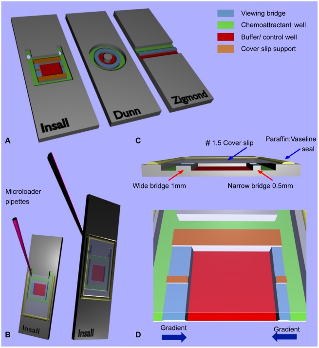
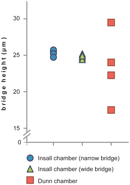
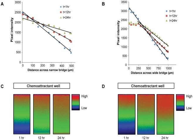
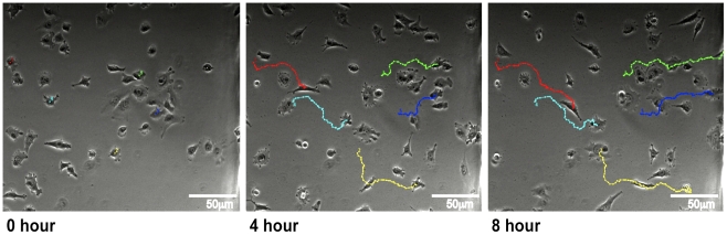
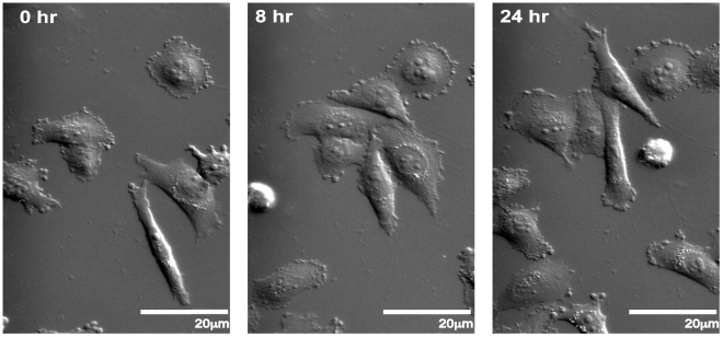
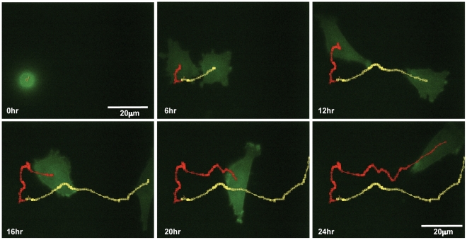
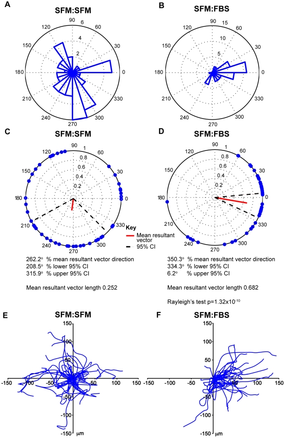
There has been a growing appreciation over the last decade that chemotaxis plays an important role in cancer migration, invasion and metastasis. Research into the field of cancer cell chemotaxis is still in its infancy and traditional investigative tools have been developed with other cell types and purposes in mind. Direct visualisation chambers are considered the gold standard for investigating the behaviour of cells migrating in a chemotactic gradient. We therefore drew up a list of key attributes that a chemotaxis chamber should have for investigating cancer cell chemotaxis. These include (1) compatibility with thin cover slips for optimal optical properties and to allow use of high numerical aperture (NA) oil immersion objectives; (2) gradients that are relatively stable for at least 24 hours due to the slow migration of cancer cells; (3) gradients of different steepnesses in a single experiment, with defined, consistent directions to avoid the need for complicated analysis; and (4) simple handling and disposability for use with medical samples. Here we describe and characterise the Insall chamber, a novel direct visualisation chamber. We use it to show GFP-lifeact transfected MV3 melanoma cells chemotaxing using a 60x high NA oil immersion objective, which cannot usually be done with other chemotaxis chambers. Linear gradients gave very efficient chemotaxis, contradicting earlier results suggesting that only polynomial gradients were effective. In conclusion, the chamber satisfies our design criteria, most importantly allowing high NA oil immersion microscopy to track chemotaxing cancer cells in detail over 24 hours.


Conflict of interest statement

Competing Interests: The authors have declared that no competing interests exist.

Figures

Figure 1
Figure 1. Comparison of bridge chamber features.
(A) Schematic showing the Insall, Dunn and Zigmond chambers. The chemoattractant and buffer/control wells have been colour coded for direct comparison, along with the viewing bridges and cover slip supports. Note that the central cylindrical block on the Dunn chamber is the same height as the bridge and therefore offers no support to the cover slip. Fig 1 (B) demonstrates the versatility of the chamber with front or reverse side chemoattractant loading with no requirement for metal clips, unlike the Zigmond chamber. The latter technique involves loading after the cover slip has been secured and sealed in place with a 1∶1 mix of vaseline: paraffin, producing a tight seal that reduces the risk of evaporation during experiments over several hours. Fig 1 (C) Cross section through the Insall chamber highlighting one key feature – the ability to use thin (#1.5, 0.16–0.18 mm) cover slips that permit high NA oil immersion microscopy. Bridges of differing widths provide different gradient steepnesses. Fig (D) provides a close-up of the Insall chamber and demonstrates the directions of the two chemotactic gradients, which are unidirectional across each bridge.
Figure 2
Figure 2. Bridge heights in Insall and Dunn chambers during use.
Bridge heights are measured in µm at 4 locations across the Insall chamber narrow and wide bridges and the Dunn chamber using a #1.5, 0.16–0.18 mm cover slip. Chambers were filled with fluorescein and images were taken in a z-stack using 1 µm intervals to calculate the distance between the bridge and cover slip. The bridge height was consistent at all sites in the Insall chamber with less than 1 µm variation demonstrating that thin cover slips can be used without affecting the geometry of the chamber and hence the diffusion profile.
Figure 3
Figure 3. Stability of linear gradients.
Gradients were established for between 1 and 24 hours across the (A) narrow and (B) wide bridges in the Insall chamber. The outer chemoattractant chamber was loaded with 100 µM fluorescein in 100 mM Tris·Cl pH 8.0 and the inner control chamber with 100 mM Tris·Cl pH 8.0. Confocal images were acquired at various intervals and quantification of the gradient was performed by measuring pixel intensity from the fluoresence signal as it diffused across the (C) narrow and (D) wide bridges.
Figure 4
Figure 4. Insall chamber chemotaxis assay.
Figure shows chemotaxis of MV3 melanoma cells towards a 10% FBS gradient on the right of each image and imaged using inverted phase contrast microscopy with a 10x phase objective with time-lapse images taken every 5 minutes for 8 hours. Cell tracking lines have been created using the ImageJ MTrackJ plugin.
Figure 5
Figure 5. DIC imaging of melanoma chemotaxis.
MV3 melanoma cells migrating towards a 10% FBS chemoattractant at the top of the image(s) over the course of 24 hours were imaged in differential image contrast using a 40x 1.3NA objective.
Figure 6
Figure 6. Live cell fluorescence microscopy using high-NA objective.
Insall chamber chemotaxis assay with identical set-up and analysis to experiment in Fig. 3, but the MV3 cells are stably transfected with GFP-Lifeact. Time-lapse images are taken every 5 minutes for 24 hours with and imaged with a 60x, 1.4NA oil immersion objective. Two daughter cells can be seen emerging from mitosis, polarising and then chemotaxing towards the FBS gradient. Their paths are highlighted by the red and yellow tracking lines.
Figure 7
Figure 7. Quantification and statistical analysis.
(A, C, E) represent the results for the migration of all 46 MV3 cells tracked in a control experiment (SFM:SFM) and (B, D, F) represent the results for the migration of all 43 MV3 cells in the presence of a chemoattractant (SFM:FBS) both across the narrow bridge over 12 hours. In the chemoattractant experiment the chemoattractant is to the right. Rose plots (A & B) demonstrate that cells migrated in nearly all directions in the control experiment but predominantly up the gradient in the chemoattractant experiment. The polar plots for the control experiment (C) reveal a short mean resultant vector for the control experiment with a wide 95% confidence interval not towards the gradient. Conversely, the polar plots for the chemoattractant experiment (D) demonstrate a much longer mean resultant vector with corresponding narrow 95% confidence intervals in the direction of the chemoattractant. The evidence for directed migration is further supported by a highly significant Rayleigh test (p = 1.32×10⁻¹⁰) demonstrating a unimodal deviation from uniformity or random migration in this instance. The paths of individual cells in the spider plots highlight a strong bias for migration towards the chemoattractant.

References

    1. Pinner S, Sahai E. PDK1 regulates cancer cell motility by antagonising inhibition of ROCK1 by RhoE. Nat Cell Biol. 2008;10:127–137. - PubMed
    1. Wells A. Tumor invasion: role of growth factor-induced cell motility. Adv Cancer Res. 2000;78:31–101. - PubMed
    1. Insall RH. Understanding eukaryotic chemotaxis: a pseudopod-centred view. Nat Rev Mol Cell Biol. 2010;11:453–458. - PubMed
    1. Andrew N, Insall RH. Chemotaxis in shallow gradients is mediated independently of PtdIns 3-kinase by biased choices between random protrusions. Nat Cell Biol. 2007;9:193–200. - PubMed
    1. Van Haastert PJ, Devreotes PN. Chemotaxis: signalling the way forward. Nat Rev Mol Cell Biol. 2004;5:626–634. - PubMed

Publication types