. 2010 Dec 14;5(12):e15315.
doi: 10.1371/journal.pone.0015315.

Coxiella burnetii, the agent of Q fever, replicates within trophoblasts and induces a unique transcriptional response

Affiliations

Coxiella burnetii, the agent of Q fever, replicates within trophoblasts and induces a unique transcriptional response

Amira Ben Amara et al. PLoS One. .

Abstract

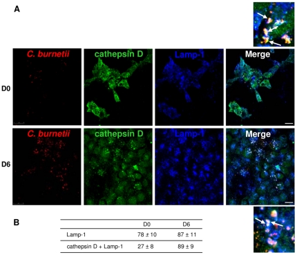
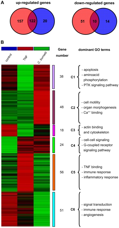
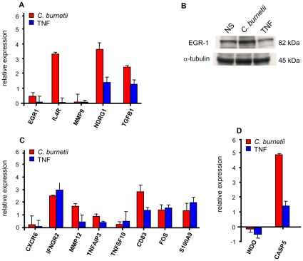
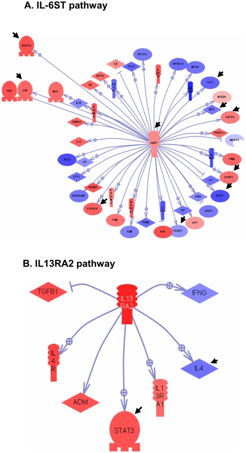
Q fever is a zoonosis caused by Coxiella burnetii, an obligate intracellular bacterium typically found in myeloid cells. The infection is a source of severe obstetrical complications in humans and cattle and can undergo chronic evolution in a minority of pregnant women. Because C. burnetii is found in the placentas of aborted fetuses, we investigated the possibility that it could infect trophoblasts. Here, we show that C. burnetii infected and replicated in BeWo trophoblasts within phagolysosomes. Using pangenomic microarrays, we found that C. burnetii induced a specific transcriptomic program. This program was associated with the modulation of inflammatory responses that were shared with inflammatory agonists, such as TNF, and more specific responses involving genes related to pregnancy development, including EGR-1 and NDGR1. In addition, C. burnetii stimulated gene networks organized around the IL-6 and IL-13 pathways, which both modulate STAT3. Taken together, these results revealed that trophoblasts represent a protective niche for C. burnetii. The activation program induced by C. burnetii in trophoblasts may allow bacterial replication but seems unable to interfere with the development of normal pregnancy. Such pathophysiologocal processes should require the activation of immune placental cells associated with trophoblasts.


Conflict of interest statement

Competing Interests: The authors have declared that no competing interests exist.

Figures

Figure 1
Figure 1. Intracellular fate of C. burnetii.
BeWo trophoblasts were incubated with different concentrations of C. burnetii (10, 50 and 200 bacteria/cell) for 4 h and extensively washed to remove free organisms (insets). They were then cultured for 9 d. The number of bacterial DNA copies was determined using qPCR. The results represent the mean ± SEM of 3 experiments.
Figure 2
Figure 2. Intracellular localization of C. burnetii.
Infected BeWo cells were labeled with anti-C. burnetii (Alexa 546), anti-cathepsin D (Alexa 488) and anti-Lamp-1 (Alexa 647) Abs and analyzed by laser scanning confocal microscopy. A, The co-localization of C. burnetii (red) with Lamp-1 (blue) and cathepsin D (green) is demonstrated by merging the respective fluorescent images. The co-localization of bacteria with both Lamp-1 and cathepsin D appears as white (see arrows), whereas the co-localization of bacteria with only Lamp-1 appears as yellow (see arrows). B, The percentage of bacteria that co-localized with Lamp-1 and cathepsin D was recorded. The results represent the mean ± SD of approximately 60 cells for each experimental condition. Scale bars represent 5 µm. D0 and D6 are day 0 and day 6 post-infection, respectively.
Figure 3
Figure 3. Venn diagram and hierarchical clustering analysis.
BeWo cells were incubated with C. burnetii (200 bacteria/cell) or TNF (10 ng/mL) for 6 h. Total RNA was extracted and, after cyanin-3 incorporation, hybridized on chips representing 31,054 genes. Only genes that were expressed with a P value <0.05 and a confidence value >0.3 in at least one condition were included in the analysis. A, The number of genes modulated in response to C. burnetii (red) and TNF (blue) stimulation is indicated. Intersections showed that 122 and 10 genes were up-regulated and down-regulated, respectively, by both C. burnetii and TNF stimulation. B, Data were analyzed using hierarchical clustering and are represented by a color gradient (Z-score) ranging from blue (down-modulation) to yellow (up-regulation). Left: unstimulated cells (NS); middle: TNF-stimulated cells; Right: C. burnetii-stimulated cells. The GO analysis was performed using two filters: 1. GO term level 2< gene <11; 2. when GO term is represented by more than 3 genes on the chip and contains more than 2 genes of the study set. The number of genes in every biological process is presented.
Figure 4
Figure 4. Real time RT-PCR and western blot analysis.
BeWo cells were stimulated with C. burnetii for 6 h. A, C and D; RNA was extracted and real time RT-PCR was performed. Results expressed in relative expression (stimulated vs. unstimulated conditions) represent the mean ± SEM of 3 independent experiments. B, Western blot analysis was performed using specific mAbs for α-tubulin and EGR-1, and bands were revealed by chemoluminescence. Their intensity was determined by densitometry and represented the mean of three experiments.
Figure 5
Figure 5. IL-6ST and IL-13RA2 pathways in C. burnetii-stimulated BeWo cells.
The IL-6ST (A) and IL-13RA2 (B) pathways induced in BeWo cells by C. burnetii stimulation were identified using Pathway Studio© software. Up-regulated genes appeared in red and down-regulated genes in blue. Arrowheads represent the differences with TNF stimulation.

Similar articles

Cited by

References

    1. Raoult D, Marrie T, Mege JL. Natural history and pathophysiology of Q fever. Lancet Infect Dis. 2005;5:219–226. - PubMed
    1. Carcopino X, Raoult D, Bretelle F, Boubli L, Stein A. Managing Q fever during pregnancy: the benefits of long-term cotrimoxazole therapy. Clin Infect Dis. 2007;45:548–555. - PubMed
    1. Stein A, Lepidi H, Mege JL, Marrie TJ, Raoult D. Repeated pregnancies in BALB/c mice infected with Coxiella burnetii cause disseminated infection, resulting in stillbirth and endocarditis. J Infect Dis. 2000;181:188–194. - PubMed
    1. Lang G, Waltner-Toews D, Menzies P. The seroprevalence of coxiellosis (Q fever) in Ontario sheep flocks. Can J Vet Res. 1991;55:139–142. - PMC - PubMed
    1. Arricau-Bouvery N, Rodolakis A. Is Q fever an emerging or re-emerging zoonosis? Vet Res. 2005;36:327–349. - PubMed

Publication types