. 2010 Dec 14;5(12):e15668.
doi: 10.1371/journal.pone.0015668.

Dispersal of biofilms by secreted, matrix degrading, bacterial DNase

Affiliations

Dispersal of biofilms by secreted, matrix degrading, bacterial DNase

Reindert Nijland et al. PLoS One. .

Abstract

Microbial biofilms are composed of a hydrated matrix of biopolymers including polypeptides, polysaccharides and nucleic acids and act as a protective barrier and microenvironment for the inhabiting microbes. While studying marine biofilms, we observed that supernatant produced by a marine isolate of Bacillus licheniformis was capable of dispersing bacterial biofilms. We investigated the source of this activity and identified the active compound as an extracellular DNase (NucB). We have shown that this enzyme rapidly breaks up the biofilms of both Gram-positive and Gram-negative bacteria. We demonstrate that bacteria can use secreted nucleases as an elegant strategy to disperse established biofilms and to prevent de novo formation of biofilms of competitors. DNA therefore plays an important dynamic role as a reversible structural adhesin within the biofilm.


Conflict of interest statement

Competing Interests: Based on these findings a UK-patent application has been filed (Burgess JG, Hall MJ, Nijland R. "Compounds and methods for biofilm disruption and prevention" GB1002396.8, 12 February, 2010.) This does not alter the authors' adherence to all the PLoS ONE policies on sharing data and materials.

Figures

Figure 1
Figure 1. Dispersal of several bacterial species by AMS supernatant.
Typical examples of dispersal of several 26 hour grown biofilm forming strains by AMS supernatant. Remaining biofilm visualised by CV staining after 30 minutes incubation with dispersal compound. x = control (only medium added), 10% = 10% of AMS supernatant added, 5% = 5% of AMS supernatant added.
Figure 2
Figure 2. Dispersal efficiency in time and concentration.
Efficiency of dispersal of B. licheniformis DSM13 24 hour old biofilm by AMS supernatant (sup.) visualised as remaining CV stain as measured by plate reader. Incubation time in minutes (') and seconds (”) indicated on the left, concentration of AMS supernatant indicated on top. The biofilm remaining is indicated with both a colour scale (dark blue: no dispersal, white: full dispersal) and as a percentage of non-dispersed biofilm (red numbers).
Figure 3
Figure 3. Efficiency of different AMS supernatant fractionation methods.
A: total supernatant of the AMS culture; B: active fraction obtained after rotary evaporation followed by Sephadex G50 gel filtration; C: active fraction obtained after freeze-drying by Sephadex-LH20 gel filtration, D+E: Active fraction obtained by TCA precipitation followed by Superose 12 gel filtration. m = Invitrogen Novex Sharp Pre-stained Marker, band sizes indicated in kDa. Arrows indicate bands 1, 2 and 3 cut out for peptide mass fingerprinting, as described in text.
Figure 4
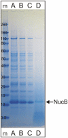
Figure 4. Heterologous overexpression of NucB in B. subtilis NZ8900.
Lane m = Invitrogen Novex Sharp Pre-stained Marker, band sizes indicated in kDa. Lanes A–C: 20 fold concentrated TCA precipitated supernatant of strain B. subtilis NZ8900+pNZ8901-nucB, loaded 20 µl (A), 10 µl (B), 5 µl (C). Lane D: loaded 20 µl unprocessed supernatant. Arrow indicates NucB position.
Figure 5
Figure 5. Comparison between NucB and DNaseI mediated biofilm dispersal.
Efficiency of dispersal of 24 hour old B. licheniformis DSM13 biofilms by the tested nucleases in decreasing concentrations. Dispersal of the target biofilm was determined using a 96 well microtitre plate setup, using a concentration range of either B. subtilis supernatant containing NucB or commercially available DNaseI. For every data point, the average of at least 6 independent wells was taken, and the experiment was repeated three times.

References

    1. Fletcher M. Bacterial biofilms and biofouling. Curr Opin Biotechnol. 1994;5:302–306. - PubMed
    1. Costerton JW, Stewart PS, Greenberg EP. Bacterial biofilms: a common cause of persistent infections. Science. 1999;284:1318–1322. - PubMed
    1. Costerton JW. Overview of microbial biofilms. J Ind Microbiol. 1995;15:137–140. - PubMed
    1. Allegrucci M, Hu FZ, Shen K, Hayes J, Ehrlich GD, et al. Phenotypic characterization of Streptococcus pneumoniae biofilm development. J Bacteriol. 2006;188:2325–2335. - PMC - PubMed
    1. Moorthy S, Watnick PI. Identification of novel stage-specific genetic requirements through whole genome transcription profiling of Vibrio cholerae biofilm development. Mol Microbiol. 2005;57:1623–1635. - PMC - PubMed

Publication types

LinkOut - more resources