. 2008 Jul;1(1):5-12.
doi: 10.1177/1756285608093978.

Treatment of neuromyelitis optica: current debate

Affiliations

Treatment of neuromyelitis optica: current debate

Tomoko Okamoto et al. Ther Adv Neurol Disord. 2008 Jul.

Abstract

Neuromyelitis optica (NMO) is an inflammatory demyelinating disease that largely affects optic nerves and spinal cord. Recent studies have identified an elevation of serum anti-aquaporin 4 antibody as a hallmark of NMO. Typical cases of NMO significantly differ from multiple sclerosis (MS) in immunological markers, histopathology, and responses to therapy. In fact, plasma exchange may be more efficacious for NMO than MS, whereas interferon-ß is recommended for MS but not for NMO. An emerging idea that pathogenesis of NMO may involve an interaction of the newly identified helper T cell subset, Th17, with B cells offers potential targets of therapy.

Keywords: Th17 cells; anti-aquaporin-4 antibody; interferon-β; multiple sclerosis; neuromyelitis optica.


Figures

Figure 1.
Figure 1.
Longitudinally extensive spinal cord lesion (LESL) in a case of NMO T2-weighted cervical MRI demonstrates an extension of T2 high density involving central gray matter, which is characteristic of LESL associated with NMO.
Figure 2.
Figure 2.
Development of large white matter lesions in a case of neuromyelitis optica (NMO) 2 months after starting interferon-/? This young female patient was aquaporin 4 antibody-positive and showed a clinical and radiological picture characteristic of NMO. However, two months after starting interferon-/? 1b treatment, she developed signs of brain hemispheres and MRI showed multiple large white matter lesions.
Figure 3.
Figure 3.
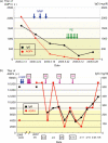
Treatment of NMO with plasmapheresis: representative cases (a) This 36-year-old female developed dysesthesia of the right leg and a constrictive band sensation in the chest region. A few days later, she experienced high fever, the loss of visual perception, progressive muscle weakness, and severe disturbance of sensation in all the limbs. She could not stand and suffered from neurogenic bladder. Treatment was initiated by the administration of 1000 mg/day of methylprednisolone (IVMP) for three consecutive days; this was followed by plasma exchange (PE) therapy which was conducted seven times over a two-week-period. The treatment was judged successful by clinical improvement as well as reduction of anti-aquaporin 4 (AQP4) antibody. (b) This 54-year-old female became completely paraplegic and was confined to bed after the development of thoracic transverse myelitis in December 2006. Although IVMP (1000 mg/day for five days followed by 500 mg/day for three days) and immunoadsorption (IA) therapies (four times) were applied, anti-AQP4 titers were somewhat elevated. So we checked the anti-AQP4 titer and total IgG before and after each of successive IA sessions. IA effectively removed the antibody and reduced the IgG amount after every IA session. But the titer and IgG returned rapidly. The anti-AQP4 antibody exhibits a higher rate of return to the basal level than that of the serum IgG. On evaluation on one month after the last IA, the patient's clinical improvement was very limited, and the anti-AQP4 antibody titer returned to the level of before starting the treatment.
Figure 4.
Figure 4.
A patient with NMO who did not respond to interferon-/? (IFN-/3) but to azathioprine. Interferon-/? was introduced to this female patient with NMO, as the patient's condition became active. However, there was no noticeable clinical benefit. After adding azathioprine, the patient entered a good remission state without any signs of relapses. Subsequently, we have withdrawn interferon-/?, and the remission state is still continuing.

References

    1. Acosta-Rodriguez E., Napolitani G., Lanzavecchia A., Sallusto F.(2007) Interleukin 1beta and 6 but not transforming growth factor-beta are essential for the differentiation of interleukin 17-producing human T helper cells. Nat Immunol 8: 942–949 - PubMed
    1. Allbutt T.(1870) On the ophthalmoscopic signs of spinal disease. Lancet 1: 76–78
    1. Baccala R., Kono D.H., Theofilopoulos A.N.(2005) Interferons as pathogenic effectors in autoimmunity. Immunol Rev 204: 9–26 - PubMed
    1. Baechler E.C., Batliwalla F.M., Karypis G., Gaffney P.M., Ortmann W.A., Espe K.J.et al.(2003) Inerferon-inducible gene expression signature in peripheral blood cells of patients with severe lupus. Proc Natl Acad Sci USA 100: 2610–2615 - PMC - PubMed
    1. Bakker J., Metz L.(2004) Devic's neuromyelitis optica treated with intravenous gamma globulin (IVIG). Can J Neurol Sci 31: 265–267 - PubMed

LinkOut - more resources