

Osteochondroplastic tracheobronchopathy: report on 02 cases and bibliographic review
- PMID: 21180949
- PMCID: PMC9443731
- DOI: 10.1590/S1808-86942010000600019
Osteochondroplastic tracheobronchopathy: report on 02 cases and bibliographic review
Abstract
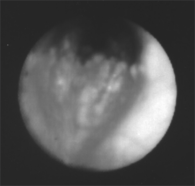
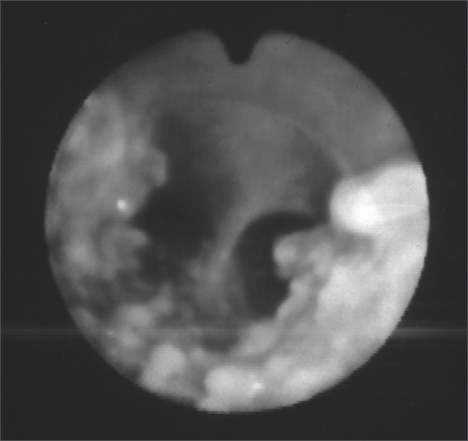
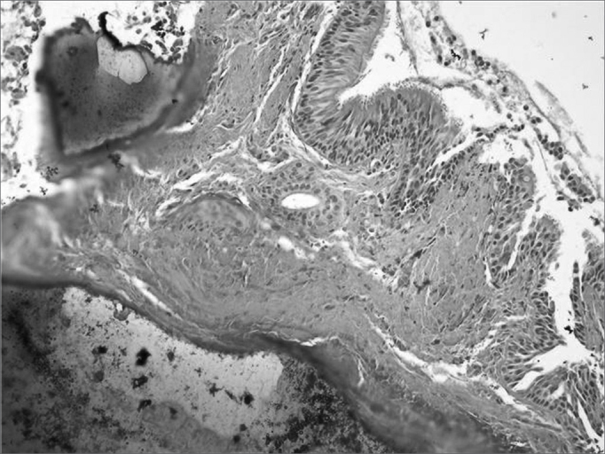
Osteochondroplastic tracheobronchopathy (OT) is a rare benign disorder of the lower part of the trachea and the upper part of the main bronchus characterized by numerous submucosal calcified nodules, sessile, cartilaginous and/or osseous with laryngotracheobronchial lumen projection. There are less than 400 cases reported in the word literature.
Aim: to report and discuss 02 cases of OT with a bibliography review.
Materials and methods: we report on 02 cases with bibliography revision from MEDLINE, LILACS and PUBMED data.
Study design: observational, descriptive, case reports.
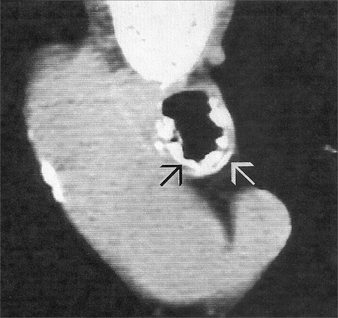
Conclusion: the symptoms result from airway obstruction, causing dry cough, dyspnea and recurrent respiratory tract infections. The diagnostic hypothesis is established by endoscopy of the upper airway (laryngo-tracheo-bronchoscopy), and the trachea/chest computed tomography is the best image exam to define tracheal nodule alterations. The differential diagnoses are papillomatosis, amyloidosis and sarcoidosis chondrosarcoma hamartoma and calcified paratracheal lymph nodes. There is no specific treatment and the prognosis is good. Surgery is restricted to moderate or severe airway obstructions. Otorhinolaryngologists must include OT in the differential diagnosis of cases of upper airway and tracheobronchial tree suggestive symptoms.
Figures
References
-
- Tabilo FP. Traqueobroncopatia Osteoplástica. Enferm Respir Cir Torac. 1987;3:206–209.
-
- Birzgalis AR, Farrington WT, O'keefe L, Shaw J. Localized tracheopathia osteoplastica of the subglottis. J Laryngol Otol. 1993;107:352–353. - PubMed
-
- Sarmiento SA, Álvarez MCB, Delgado FA. Traqueobroncopatía Osteocondroplásica. Rev Cuba Oncol. 2000;2(16):88–92.
-
- Shigematsu Y, Sugio K, Yasuda M, Sugaya M, Ono K, Takenoyama M, et al. Tracheobronchopathia Osteochondroplastica Occurring in a Subsegmental Bronchus and Causing Obstructive Pneumonia. Ann Thorac Surg. 2005;80:1936–1938. - PubMed
Publication types
MeSH terms
LinkOut - more resources
Full Text Sources
Medical
