. 2011 Aug;19(7):582-8.
doi: 10.3109/1061186X.2010.542245. Epub 2010 Dec 24.

Targeted delivery of a proapoptotic peptide to tumors in vivo

Affiliations

Targeted delivery of a proapoptotic peptide to tumors in vivo

Sandrine Dufort et al. J Drug Target. 2011 Aug.

Abstract

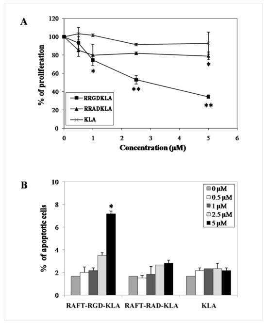
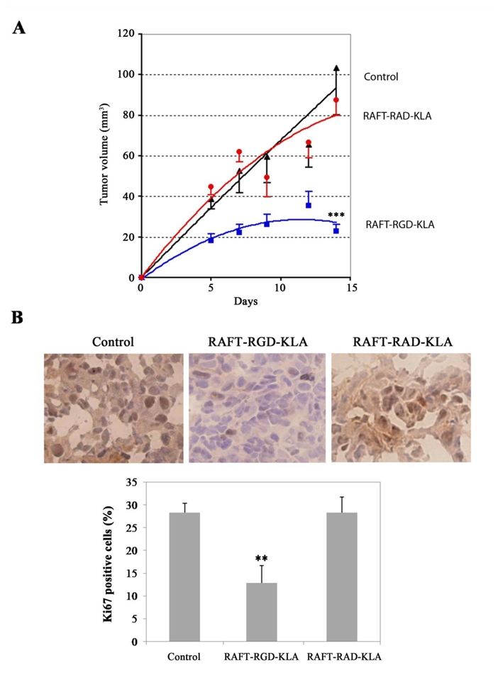
RGD peptides recognize the α(v)β(3) integrin, a receptor that is overexpressed on the surface of both tumor blood vessels and cancerous cells. These peptides are powerful tools that act as single antiangiogenic molecules, but recently also have been used for tumor imaging and drug targeting. We designed the molecule RAFT-(c[-RGDfK-])(4), a constrained and chemically defined entity that can be produced at clinical-grade quality. This scaffold was covalently coupled via a labile bridge to the proapoptotic peptide (KLAKLAK)(2) (RAFT-RGD-KLA). A fluorescent, activatable probe was also introduced, allowing intracellular localization. At 2.5 µM, this molecule induced the intracellular release of an active KLA peptide, which in turn caused mitochondrial depolarization and cell death in vitro in tumor cells. In a mouse model, the RAFT-RGD-KLA peptide was found to prevent the growth of remote subcutaneous tumors. This study demonstrated that the antitumor peptide is capable of killing tumor cells in an RGD-dependent manner, thus lowering the nonspecific cytotoxic effects expected to occur when using cationic cytotoxic peptides. Thus, this chemistry is suitable for the design of complex, multifunctional molecules that can be used for both imaging and therapeutics, representing the next generation of perfectly controlled, targeted drug-delivery systems.


Conflict of interest statement

Declaration of Interest

All the authors declare that there is no conflict of interest

Figures

Figure 1
Figure 1
Representative picture of RAFT-(cRGD)4-QSY21®-S-S-(KLAKLAK)2-Cy5 in medium alone (A-C) or with 1 μl β-mercaptoethanol (B–D). This reducing agent induced disulfide bridge rupture and allowed the detection of the Cy5 fluorescence with the 2D-FRI imaging system before (C) and after (D) the addition of DMSO β-mercaptoethanol.
Figure 2
Figure 2. Confocal microscopy on living cells
The cells were starved for 30 min prior to the addition of 2 μM RAFT-(cRGD)4-QSY21®-S-S-(KLAKLAK)2-Cy5. After 1 hr, the mitochondria were labelled with Mitotracker. Confocal imaging was performed between 1 and 2 hr. Colocalization of the mitochondria (green) and (KLAKLAK)2-Cy5 (red) appears yellow. Note that the red fluorescence was not observed under excitation at 633 nm until 1 hr after addition of RAFT-(cRGD)4-QSY21®-S-S-(KLAKLAK)2-Cy5 in the medium. Pictures are representative of cells observed on three separate plates. Scale bar: 10 μm.
Figure 3
Figure 3. RAFT-RGD-KLA-mediated cytotoxicity
(A) IGROV-1 cells were treated for 48 hr with 0.5, 1.0, 2.5 or 5.0 μM of each compound, and the number of viable cells was counted. The results are expressed as the percentage of cells compared to untreated control. (B) The cells were then fixed, and their nuclei were labelled using Hoechst 33342. The number of apoptotic nuclei was counted. The results are expressed as the mean ± SD (n = 3). * P < 0.05, ** P < 0.01.
Figure 4
Figure 4. Mitochondrial activity in IGROV-1 cells
(A) Example of confocal imaging of polarized (red, ex. 543 nm) and depolarized (green, ex. 488 nm) mitochondria in IGROV-1 living cells before and after treatment with RAFT-RGD-KLA at a concentration of 1 μM. Corresponding 3D surface topographies from confocal imaging are represented below. Treatment with RAFT-RGD-KLA induced an increase in green fluorescence, i.e., mitochondria depolarization (p < 0.05). Topography scale: low (in blue) to high fluorescence intensity (in red). Angle of view: lower right corner. (B) Polarized to depolarized mitochondria ratio from JC-1 staining of IGROV-1 cells after 2 hr of treatment with PBS (control) or 2 μM RAFT-RGD-KLA, KLA or RAFT-RAD-KLA. RAFT-RGD-KLA was found to significantly decrease the mitochondrial ratio compared to the control condition (p = 0.002 ** and p = 0.095, respectively).
Figure 5
Figure 5. Inhibition of tumour growth in vivo by RAFT-RGD-KLA
(A) At day 0, IGROV-1 cells were implanted subcutaneously in the right legs of nude mice. Mice were treated daily for 14 days with an IP injection of 200 μl PBS (control) or with solutions containing 0.12 μmol RAFT-RGD-KLA or RAFT-RAD-KLA. Tumour volumes are expressed as the mean tumour volume ± S.E.M. (n = 5–7 per group; *** P < 0.0001). (B) Ki67 nuclear protein detected by immunostaining on frozen tumour sections from control mice, mice treated by RAFT-RGD-KLA or RAFT-RAD-KLA, as indicated. Histogram: the percentage of positive cells for Ki67 was determined after counting the number of positively stained cells per 1,000 cells on the tissue section. The results are expressed as the mean ± SD; ** P < 0.01 for comparisons between RAFT-RGD-KLA and control.

References

    1. Ahmadi M, Sancey L, Briat A, Riou L, Boturyn D, Dumy P, Fagret D, Ghezzi C, Vuillez JP. Chemical and Biological Evaluations of an (111) In-Labeled RGD-Peptide Targeting Integrin Alpha(V) Beta(3) in a Preclinical Tumor Model. Cancer Biother Radiopharm. 2008;23:691–700. - PubMed
    1. Borgne-Sanchez A, Dupont S, Langonne A, Baux L, Lecoeur H, Chauvier D, Lassalle M, Deas O, Briere JJ, Brabant M, Roux P, Pechoux C, Briand JP, Hoebeke J, Deniaud A, Brenner C, Rustin P, Edelman L, Rebouillat D, Jacotot E. Targeted Vpr-derived peptides reach mitochondria to induce apoptosis of alphaVbeta3-expressing endothelial cells. Cell Death Differ. 2007;14:422–435. - PubMed
    1. Boturyn D, Coll JL, Garanger E, Favrot MC, Dumy P. Template assembled cyclopeptides as multimeric system for integrin targeting and endocytosis. J Am Chem Soc. 2004;126:5730–5739. - PubMed
    1. Chen J, Xu XM, Underhill CB, Yang S, Wang L, Chen Y, Hong S, Creswell K, Zhang L. Tachyplesin activates the classic complement pathway to kill tumor cells. Cancer Res. 2001;65:4614–4622. - PubMed
    1. Chen Y, Xu X, Hong S, Chen J, Liu N, Underhill CB, Creswell K, Zhang L. RGD-Tachyplesin inhibits tumor growth. Cancer Res. 2001;61:2434–2438. - PubMed

Publication types