

Comment
doi: 10.1016/j.cell.2010.12.005.
Duchenne muscular dystrophy models show their age
Affiliations
- PMID: 21183068
- PMCID: PMC3038548
- DOI: 10.1016/j.cell.2010.12.005
Item in Clipboard
Comment
Duchenne muscular dystrophy models show their age
Cell.
.
Abstract
The lack of appropriate animal models has hampered efforts to develop therapies for Duchenne muscular dystrophy (DMD). A new mouse model lacking both dystrophin and telomerase (Sacco et al., 2010) closely mimics the pathological progression of human DMD and shows that muscle stem cell activity is a key determinant of disease severity.
Copyright © 2010 Elsevier Inc. All rights reserved.
Figures
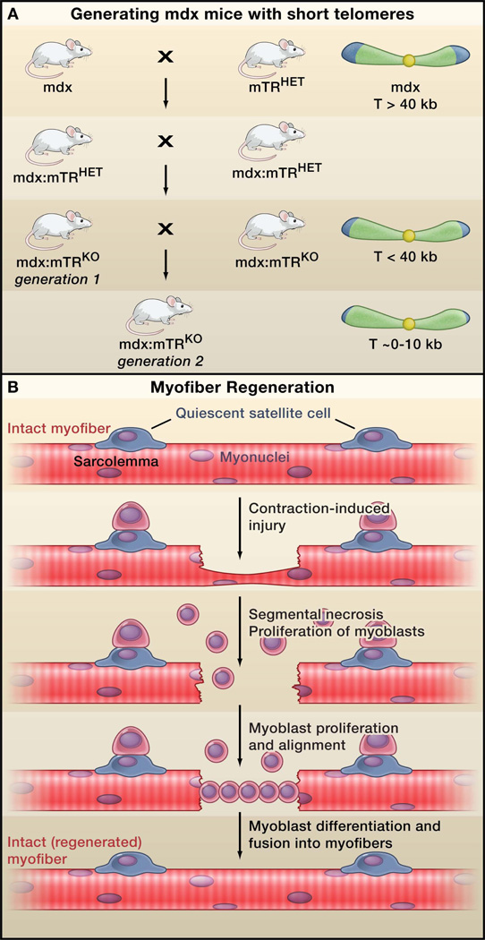
New model for Duchenne muscular dystrophy (DMD). (A) Sacco et al. (2010) generate a new mouse model for DMD by crossing dystrophin-deficient mdx mice (the current DMD model) with mice carrying a mutation in the telomerase gene (mTRHET mice). Inbreeding of the resulting heterozygotes (mdx/mTRHET) produces double knockout mice (mdx/mTRKO) that show decreasing telomere length (T) in the first and second generations, and a corresponding decrease in the capacity of myogenic stem cells (satellite cells) to proliferate. These mice display a rapidly progressing muscular dystrophy phenotype that more closely mimics the human disease compared to the current model. Note that mdx is an X-linked gene. For simplicity, the complete mdx genotype is not depicted in the figure. (B) Dystrophic muscles are highly susceptible to contraction-induced injury, which impairs the protective sheath around myofibers (the sarcolemma), leading to myofiber necrosis and activation of nearby satellite cells. Activated satellite cells divide asymmetrically, giving rise to one satellite cell, and a proliferating daughter cell that generates myoblasts. The myoblasts migrate to the site of injury, differentiate into myocytes, align and fuse into the damaged myofiber, repairing the lesion.
Comment on
-
Short telomeres and stem cell exhaustion model Duchenne muscular dystrophy in mdx/mTR mice.Cell. 2010 Dec 23;143(7):1059-71. doi: 10.1016/j.cell.2010.11.039. Epub 2010 Dec 9. Cell. 2010. PMID: 21145579 Free PMC article.
References
-
- Blasco MA, Lee HW, Hande MP, Samper E, Lansdorp PM, DePinho RA, Greider CW. Cell. 1997;91:25–34. - PubMed
-
- Chamberlain JS, Metzger J, Reyes M, Townsend D, Faulkner JA. FASEB J. 2007;21:2195–2204. - PubMed
-
- Collins CA, Olsen I, Zammit PS, Heslop L, Petrie A, Partridge TA, Morgan JE. Cell. 2005;122:289–301. - PubMed
-
- Deconinck AE, Rafael JA, Skinner JA, Brown SC, Potter AC, Metzinger L, Watt DJ, Dickson JG, Tinsley JM, Davies KE. Cell. 1997;90:717–727. - PubMed
Publication types
Grants and funding
LinkOut - more resources
Full Text Sources
Molecular Biology Databases
