. 2010 Dec 23;143(7):1072-83.
doi: 10.1016/j.cell.2010.11.036.

C/EBPβ controls exercise-induced cardiac growth and protects against pathological cardiac remodeling

Affiliations

C/EBPβ controls exercise-induced cardiac growth and protects against pathological cardiac remodeling

Pontus Boström et al. Cell. .

Abstract

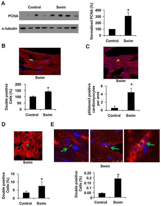
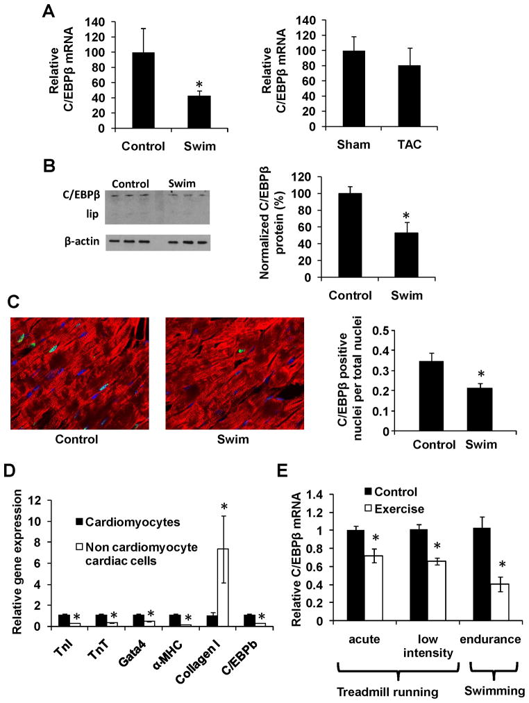
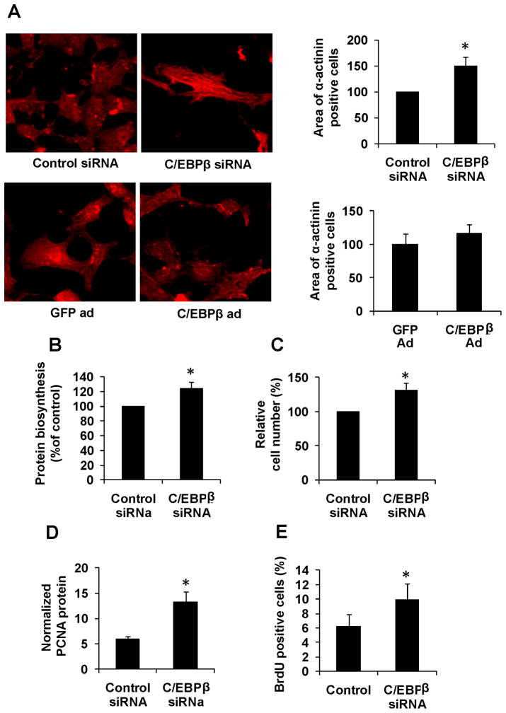
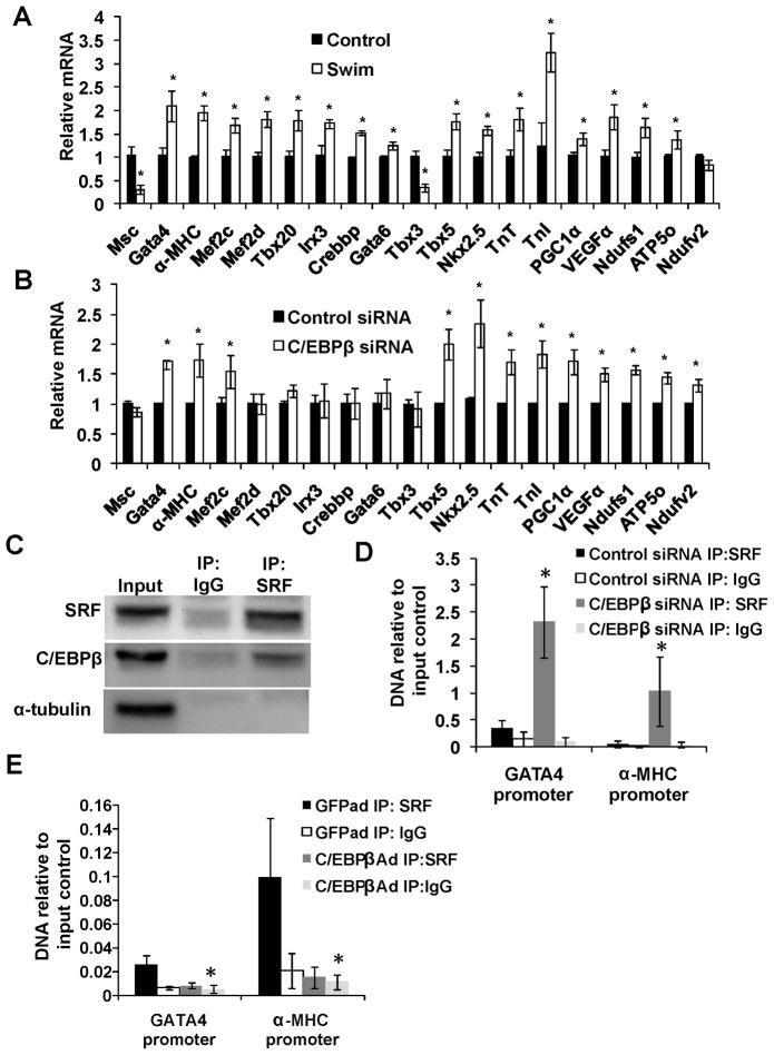
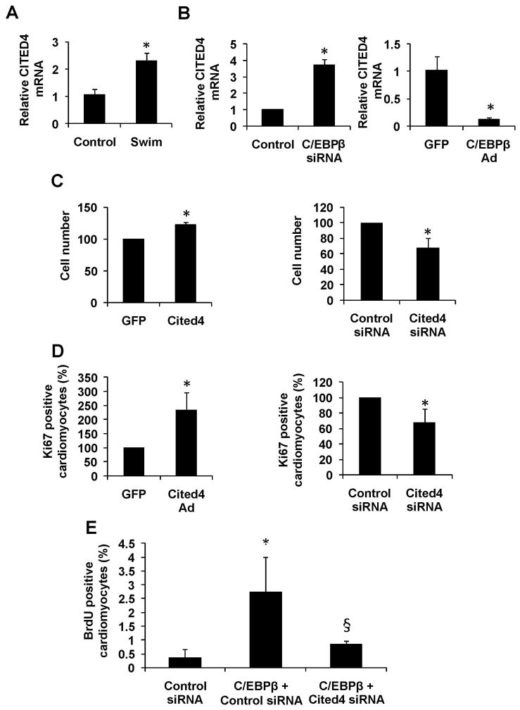
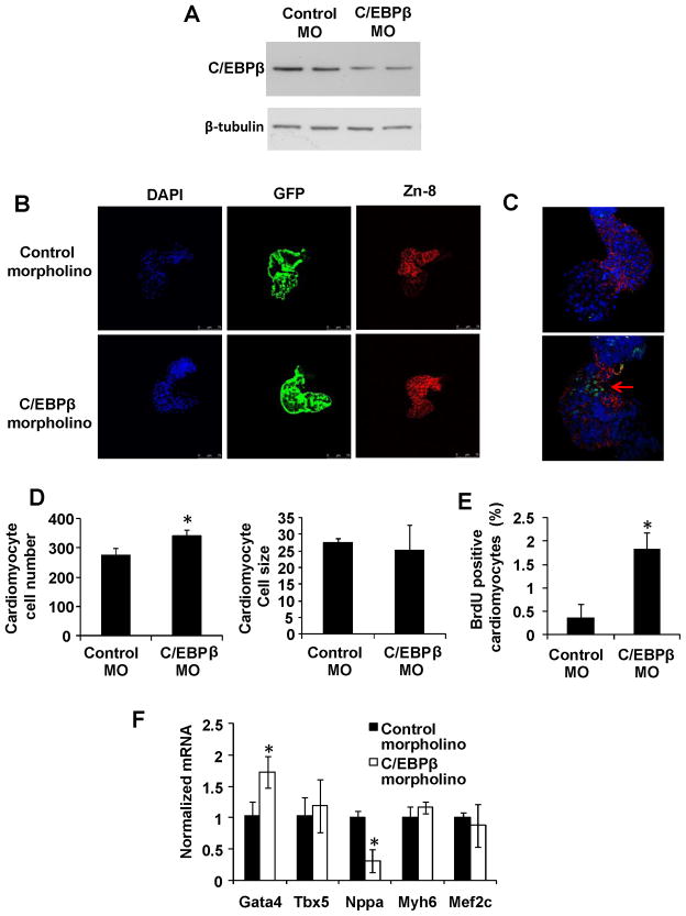
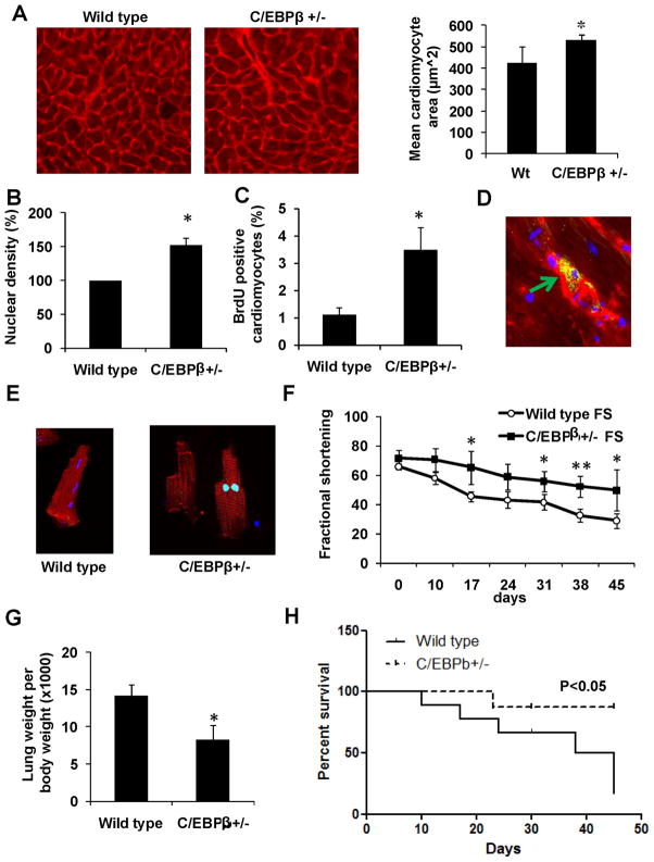
The heart has the ability to grow in size in response to exercise, but little is known about the transcriptional mechanisms underlying physiological hypertrophy. Adult cardiomyocytes have also recently been proven to hold the potential for proliferation, a process that could be of great importance for regenerative medicine. Using a unique RT-PCR-based screen against all transcriptional components, we showed that C/EBPβ was downregulated with exercise, whereas the expression of CITED4 was increased. Reduction of C/EBPβ in vitro and in vivo resulted in a phenocopy of endurance exercise with cardiomyocyte hypertrophy and proliferation. This proliferation was mediated, at least in part, by the increased CITED4. Importantly, mice with reduced cardiac C/EBPβ levels displayed substantial resistance to cardiac failure upon pressure overload. These data indicate that C/EBPβ represses cardiomyocyte growth and proliferation in the adult mammalian heart and that reduction in C/EBPβ is a central signal in physiologic hypertrophy and proliferation.


Figures

Figure 1
Figure 1. A swim model induces physiological cardiac hypertrophy and cardiomyocyte proliferation
A. Mice swam using a ramp protocol for 2 weeks with respective controls (n=4), and were assayed for PCNA protein levels with subsequent quantification. Data is presented as PCNA per α-tubulin after subtraction of background. Data is representative for two independent mouse cohorts. B-C. Representative image and quantification of Ki67 (B) and phosphoHistone3 (C) staining from exercised mice (green) counterstained with α-actinin (red) and DAPI (blue) in cardiac tissue. D. Exercised and control mice were injected with BrdU 3 days prior to the final exercise day. The image shows a representative section from the exercised cohort. BrdU is labeled in green, α-actinin in red and DAPI in blue. E. Representative image and quantification of Aurora B kinase (green) staining from exercised mice counterstained with α-actinin (red) and DAPI (blue) in cardiac tissue. All quantifications were based on at least 80 (blinded) images from each group (from at least 4 mice). Any uncertainty concerning whether a ki67, AuroraB or BrdU positive cell was completely within an α-actinin positive area was resolved using confocal z-stacks. * indicates p<0.05 vs respective control using students t-test. Se also Supplemental Figure S1.
Figure 2
Figure 2. C/EBPβ is expressed in cardiomyocytes and down regulated with endurance exercise
A. Cardiac C/EBPβ mRNA levels in a model for physiological (swim) and pathological (TAC) hypertrophy with respective controls (n=4) as described in methods. B. Western blot analysis of C/EBPβ and β-actin in the swim cohort (above) with subsequent quantification. Data is presented as percent of control with C/EBPβ relative to β-actin after background subtraction. C. Confocal microscopy after immunohistochemistry against C/EBPβ (green), α-actinin (red) and DAPI (blue) in cardiac tissue with subsequent quantification in indicated groups. Data is presented as the ratio of C/EBPβ positive cardiomyocyte nuclei. D. Gradient fractionation of primary rat neonatal cardiac cells followed by expression analysis of cardiomyocyte and fibroblast markers. E. Cardiac C/EBPβ mRNA levels from three different exercise regimens; “acute” = 40 minutes treadmill running (n=6), “low intensity” = 2 weeks of 30 minutes daily running (n=6) and “endurance” = the swimming protocol as described in methods (n=4). * indicates p<0.05 vs respective control using students t-test. Se also Supplemental Figure S2.
Figure 3
Figure 3. Reduction of C/EBPβ in primary cardiomyocytes results in hypertrophic cell growth and proliferation
Primary rat neonatal cardiomyocytes treated with either a C/EBPβ siRNA or a C/EBPβ expressing adenovirus with respective controls. All experiments were performed 48 hours after transfection / transduction. A. Immunohistochemistry against α-actinin followed by cell area quantifications as described in methods. At least 100 cells were quantified in all groups. B. Protein biosynthesis measured as 35S-Met incorporation into the protein pool after a 1h pulse. Data is presented as percent of control. C. Quantification of cell numbers in primary after transfection with indicated siRNA constructs. D. Western blot analysis of PCNA followed by quantification (n=4). Data is presented as PCNA relative to β-actin after background subtraction. E. Primary rat neonatal cardiomyocytes were treated with BrdU 24 hours after transfection with indicated siRNA. Cells were then stained against BrdU and α-actinin and BrdU-positive cardiomyocytes were counted normalized to total number of cardiomyocytes. * indicates p<0.05 vs respective control using students t-test. Se also Supplemental Figure S3.
Figure 4
Figure 4. C/EBPβ controls a exercise –induced gene set by inhibiting SRF DNA binding
QPCR analysis of indicated genes in hearts from endurance exercised mice (A) and in primary cardiomyocytes treated with C/EBPβ or control siRNA for 48 hours (B). * indicates p<0.05 vs respective control using students t-test. C, Immunoprecipitation against SRF or a non-immune IgG in primary cardiomyocytes followed by western blot against SRF, C/EBPβ and the control protein α-tubulin as indicated. D-E. Chromatin immunoprecipitation in primary cardiomyocytes treated with either C/EBPβ siRNA (D) or C/EBPβ adenoviral over expression (E) with respective controls. Precipitated DNA was then analyzed with RT-PCR using primers directed against promoter regions in the Gata4 and myh6 gene. ChIP experiments were repeated at least three times with similar results. * indicates p<0.05 vs respective control using one-way ANOVA statistics. Se also Supplemental Figure S4.
Figure 5
Figure 5. CITED4 is up regulated with endurance exercise, regulated by C/EBPβ, and its forced expression induced cardiomyocyte proliferation in vitro
A. mRNA levels of cardiac CITED4 in control and endurance exercised (swim) mice normalized to 18S expression. B. mRNA expression of CITED4 in primary rat neonatal cardiomyocytes after siRNA knockdown (left panel) or adenoviral over expression (right panel) of CITED4. Expression levels is normalized to 18S. C-D. Rat neonatal cardiomyocytes treated with either an adenovirus over expressing CITED4 (left) or a siRNA directed against CITED4 (right). Cells were then stained against α-actinin and ki67, and positive cells were counted in 20x view fields from 15 random images per group. E. Rat neonatal cardiomyocytes treated with control siRNA, C/EBPβ siRNA + control siRNA or C/EBPβ siRNA + Cited4 siRNA, followed by assay of BrdU incorporation as previously described. Data is pooled from three experiments and presented as percent of control. * indicates p<0.05 vs respective control, and § versus C/EBPβ siRNA using one-way ANOVA statistics. Se also Supplemental Figure S5.
Figure 6
Figure 6. C/EBPβ reduction in the Zebrafish embryo results in cardiomyocyte proliferation
An anti-C/EBPβ morpholino was injected into one-cell stage cmlc-GFP transgenic Zebrafish embryos. A. Western blot analysis against indicated proteins in zebrafish embryos 48 hpf. B. Dissection of beating hearts at 36 hours followed by integrin staining (blue = DAPI, green = GFP and red = zn-8). C. 20 minutes BrdU labeling prior to dissection of beating hearts and BrdU (green) / zn-8 (red) staining. D. Quantification (B) from totally 30 hearts per group. E. Quantification of integrin and BrdU positive cardiomyocytes (C) from n=6 embryos per group. Data is presented as percent of control and representative of two independent experiments. F, RT-PCR analysis of indicated transcripts. * indicates p<0.05 vs respective control using students t-test. Se also Supplemental Figure S6.
Figure 7
Figure 7. C/EBPβ+/− mice have cardiac hypertrophy, increased proliferation markers and are resistant to pressure overload
Cardiomyocyte size was measured in cardiac tissue from wild type (n=5) and C/EBPβ+/− (n=7) mice using WGA membrane staining (A). B-C. Quantification of nuclear density assayed (B) and BrdU incorporation (C) from indicated mice. D. Typical image of Aurora B kinase (green) positive cardiomyocyte from C/EBPβ+/− mouse heart, counterstained with anti-α-actinin (red) and dapi (blue). E. Immunohistochemistry against phosphohistone H3 (green) in primary cardiomyocyte from an adult C/EBPβ+/− mouse heart counterstained with α-actinin (red) and dapi (blue). F. Wild type and C/EBPβ+/− (n=6 and 7) mice subjected to TAC and assayed for fractional shortening at indicated times. G. Lung weight 52 days after TAC intervention. H. Kaplan Meyer plot of cardiac failure free fraction at indicated times in wild type and C/EBPβ+/− mice following TAC procedure. * indicates p<0.05 vs respective control using students t-test. Se also Supplemental Figure S7.

Comment in

  • Heart-healthy hypertrophy.
    Trivedi CM, Epstein JA. Trivedi CM, et al. Cell Metab. 2011 Jan 5;13(1):3-4. doi: 10.1016/j.cmet.2010.12.012. Cell Metab. 2011. PMID: 21195341 Free PMC article.

References

    1. Akazawa H, Komuro I. Roles of cardiac transcription factors in cardiac hypertrophy. Circ Res. 2003;92:1079–1088. - PubMed
    1. Aoyama T, Matsui T, Novikov M, Park J, Hemmings B, Rosenzweig A. Serum and glucocorticoid-responsive kinase-1 regulates cardiomyocyte survival and hypertrophic response. Circulation. 2005;111:1652–1659. - PubMed
    1. Arany Z, Novikov M, Chin S, Ma Y, Rosenzweig A, Spiegelman BM. Transverse aortic constriction leads to accelerated heart failure in mice lacking PPAR-gamma coactivator 1alpha. Proc Natl Acad Sci U S A. 2006;103:10086–10091. - PMC - PubMed
    1. Bergmann O, Bhardwaj RD, Bernard S, Zdunek S, Barnabe-Heider F, Walsh S, Zupicich J, Alkass K, Buchholz BA, Druid H, et al. Evidence for cardiomyocyte renewal in humans. Science. 2009;324:98–102. - PMC - PubMed
    1. Bersell K, Arab S, Haring B, Kuhn B. Neuregulin1/ErbB4 signaling induces cardiomyocyte proliferation and repair of heart injury. Cell. 2009;138:257–270. - PubMed

Publication types

LinkOut - more resources