. 2011 May 1;69(9):857-66.
doi: 10.1016/j.biopsych.2010.09.053. Epub 2010 Dec 23.

Gray matter alterations in adults with attention-deficit/hyperactivity disorder identified by voxel based morphometry

Affiliations

Gray matter alterations in adults with attention-deficit/hyperactivity disorder identified by voxel based morphometry

Larry J Seidman et al. Biol Psychiatry. .

Abstract

Background: Gray and white matter volume deficits have been reported in many structural magnetic resonance imaging (MRI) studies of children with attention-deficit/hyperactivity disorder (ADHD); however, there is a paucity of structural MRI studies of adults with ADHD. This study used voxel based morphometry and applied an a priori region of interest approach based on our previous work, as well as from well-developed neuroanatomical theories of ADHD.

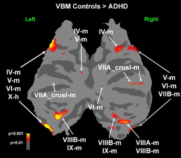
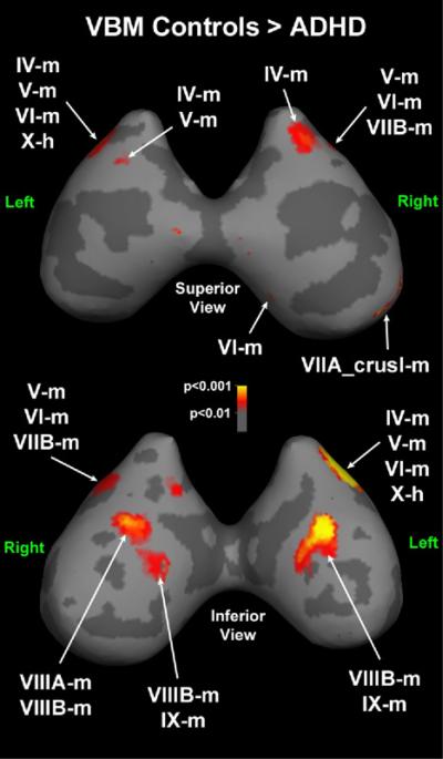
Methods: Seventy-four adults with DSM-IV ADHD and 54 healthy control subjects comparable on age, sex, race, handedness, IQ, reading achievement, frequency of learning disabilities, and whole brain volume had an MRI on a 1.5T Siemens scanner. A priori region of interest hypotheses focused on reduced volumes in ADHD in dorsolateral prefrontal cortex, anterior cingulate cortex, caudate, putamen, inferior parietal lobule, and cerebellum. Analyses were carried out by FSL-VBM 1.1.

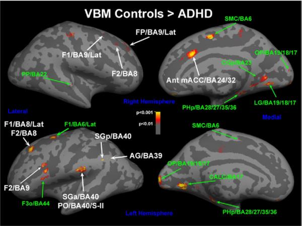
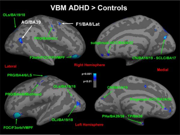
Results: Relative to control subjects, ADHD adults had significantly smaller gray matter volumes in parts of six of these regions at p ≤ .01, whereas parts of the dorsolateral prefrontal cortex and inferior parietal lobule were significantly larger in ADHD at this threshold. However, a number of other regions were smaller and larger in ADHD (especially fronto-orbital cortex) at this threshold. Only the caudate remained significantly smaller at the family-wise error rate.

Conclusions: Adults with ADHD have subtle volume reductions in the caudate and possibly other brain regions involved in attention and executive control supporting frontostriatal models of ADHD. Modest group brain volume differences are discussed in the context of the nature of the samples studied and voxel based morphometry methodology.


Figures

Figure 1
Figure 1
Voxel based morphometry control subjects > attention-deficit/hyperactivity disorder (ADHD): cortical regions that are smaller in ADHD compared with control subjects. Labels with arrows are those regions that are smaller in ADHD (p < .01) and correspond to those described in Table 2 for predicted regions of interest. Note those regions that are smaller at this threshold, which are considered exploratory findings outside of the predicted regions of interest, are in Table S1 in Supplement 1. Significant differences are displayed on the inflated surface of the Montreal Neurological Institute brain. AG, angular gyrus; Ant mACC, anterior middle cingulate cortex; BA, Brodmann area; CALC, calcarine area; CGp, posterior cingulate gyrus; F1, superior frontal gyrus; F2, middle frontal gyrus; F3o, inferior frontal gyrus, pars opercularis; FP, frontal pole; Lat, lateral; LG, lingual gyrus; OP, occipital pole; PHp, posterior parahippocampal gyrus; PO, parietal operculum; PP, planum polare; S-II, somatosensory area - II; SGa, anterior supramarginal gyrus; SGp, posterior supramarginal gyrus; SMC, supplementary motor cortex; VBM, voxel based morphometry.
Figure 2
Figure 2
Voxel based morphometry control subjects < attention-deficit/hyperactivity disorder (ADHD): cortical regions that are larger in ADHD compared with control subjects. Labels with arrows are those regions that are larger in ADHD (p < .01) and correspond to those described in Table 2 for predicted regions of interest. Note those regions that are larger at this threshold, which are considered exploratory findings, outside of the predicted regions of interest are in Table S1 in Supplement 1. Significant differences are displayed on the inflated surface of the Montreal Neurological Institute brain. ACC, anterior cingulate cortex; AG, angular gyrus; BA, Brodmann area; CGp, posterior cingulate gyrus; CN,cuneus; F1, superior frontal gyrus; F3 or b, inferior frontal gyrus, pars orbitalis; FOC, fronto-orbital cortex; FP, frontal pole; Lat, lateral; Li, lateral inferior; LS, lateral superior; OLi, lateral occipital area, inferior part; OLs, lateral occipital area, superior part; PHa, anterior parahippocampal gyrus; PO, parietal operculum; PP, planum polare; PRG, precentral gyrus; SCLC, supracalcarine cortex; TP, temporal pole; VBM, voxel based morphometry; VMPF, ventromedial prefrontal cortex.
Figure 3
Figure 3
Cerebellar regions that are smaller in attention-deficit/hyperactivity disorder compared with control subjects on a flattened brain. Labels with arrows are smaller in attention-deficit/hyperactivity disorder (p < .01) and correspond to those described in Table 2. Significant differences are displayed on the flattened cerebellar surface of the Montreal Neurological Institute brain. ADHD, attention-deficit/hyperactivity disorder; IV-m, culmen superior; IX-m, tonsil; V-m, culmen inferior; VI-m, simplex; VIIA_crusI-m, superior semilunar lobule; VIIB-m, paramedian/gracilis; VIIIA-m, biventer (pars copularis); VIIIB-m, biventer (pars paraflocculus dorsalis); X-h, hemispheric zone of lobule X; X-m, flocculus; VBM, voxel based morphometry.
Figure 4
Figure 4
Cerebellar regions that are smaller in attention-deficit/hyperactivity disorder compared with control subjects as seen on an inflated brain. Labels with arrows are smaller in attention-deficit/hyperactivity disorder (p < .01) and correspond to those described in Table 2. Significant differences are displayed on the inflated cerebellar surface of the Montreal Neurological Institute brain. ADHD, attention-deficit/hyperactivity disorder; IV-m, culmen superior; IX-m, tonsil; V-m, culmen inferior; VI-m, simplex; VIIA_crusI-m, superior semilunar lobule; VIIB-m, paramedian/gracilis; VIIIA-m, biventer (pars copularis); VIIIB-m, biventer (pars paraflocculus dorsalis); X-h, hemispheric zone of lobule X; X-m, flocculus; VBM, voxel based morphometry.

References

    1. Faraone SV, Biederman J, Spencer T, Wilens T, Seidman LJ, Mick E, Doyle A. Attention deficit hyperactivity disorder in adults: An overview. Biol Psychiatry. 2000;48:9–20. - PubMed
    1. Biederman J. Attention-deficit/hyperactivity disorder: A life span perspective. J Clin Psychiatry. 1998;59:4–16. - PubMed
    1. Biederman J, Mick E, Faraone SV. Age-dependent decline of symptoms of attention deficit hyperactivity disorder: Impact of remission definition and symptom type. Am J Psychiatry. 2000;157:816–818. - PubMed
    1. Kessler RC, Adler L, Barkley R, Biederman J, Conners CK, Demler O, et al. The prevalence and correlates of adult ADHD in the United States: Results from the national comorbidity survey replication. Am J Psychiatry. 2006;163:716–723. - PMC - PubMed
    1. Faraone SV, Biederman J. What is the prevalence of adult ADHD? Results of a population screen of 966 adults. J Atten Disord. 2005;9:384–391. - PubMed

Publication types