

First morphological characterization of 'Candidatus Mycoplasma turicensis' using electron microscopy
- PMID: 21183295
- PMCID: PMC3127424
- DOI: 10.1016/j.vetmic.2010.11.020
First morphological characterization of 'Candidatus Mycoplasma turicensis' using electron microscopy
Abstract
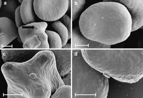
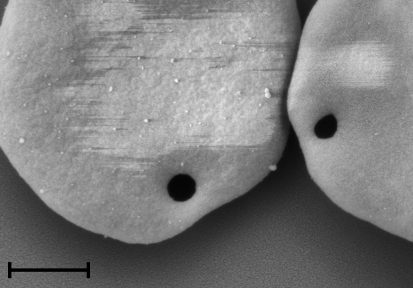
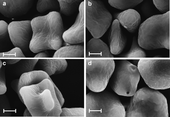
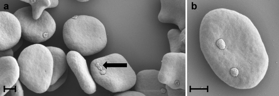
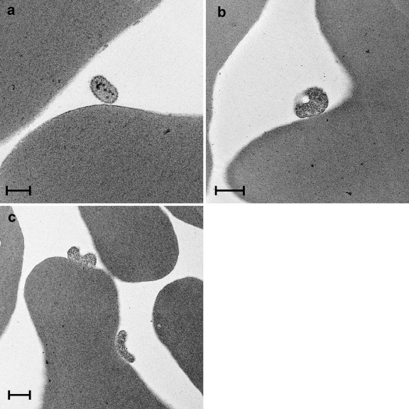
At least three haemotropic mycoplasmas have been recognized in cats: Mycoplasma haemofelis (Mhf), 'Candidatus Mycoplasma haemominutum' (CMhm) and 'Candidatus M. turicensis' (CMt). The latter was originally identified in a Swiss pet cat with haemolytic anaemia and shown to be prevalent in domestic cats and wild felids worldwide using molecular methods. So far, there has been no confirmatory morphological evidence of the existence of CMt presumably due to low blood loads during infection while CMhm has only been characterized by light microscopy with discrepant results. This study aimed to provide for the first time electron microscopic characteristics of CMt and CMhm and to compare them to Mhf. Blood samples from cats experimentally infected with CMt, CMhm and Mhf were used to determine copy numbers in blood by real-time PCR and for transmission and scanning electron microscopy. High resolution scanning electron microscopy revealed CMt and CMhm to be discoid-shaped organisms of 0.3 μm in diameter attached to red blood cells (RBCs). In transmission electron microscopy of CMt, an oval organism of about 0.25 μm with several intracellular electron dense structures was identified close to the surface of a RBC. CMhm and CMt exhibited similar morphology to Mhf but had a smaller diameter. This is the first study to provide morphological evidence of CMt thereby confirming its status as a distinct haemoplasma species, and to present electron microscopic features of CMhm.
Copyright © 2010 Elsevier B.V. All rights reserved.
Figures
Similar articles
-
Prevalence and phylogenetic analysis of haemoplasmas from cats infected with multiple species.J Microbiol Methods. 2014 Dec;107:189-96. doi: 10.1016/j.mimet.2014.10.013. J Microbiol Methods. 2014. PMID: 25447887 Free PMC article.
-
Preliminary detection of haemoplasma in Thai cat blood samples using universal primers: identifying 'Candidatus Mycoplasma haemominutum' and closely related species.J Feline Med Surg. 2025 May;27(5):1098612X251335211. doi: 10.1177/1098612X251335211. Epub 2025 May 28. J Feline Med Surg. 2025. PMID: 40433965 Free PMC article.
-
A comparison of real-time PCR and reverse line blot hybridization in detecting feline haemoplasmas of domestic cats and an analysis of risk factors associated with haemoplasma infections.BMC Vet Res. 2012 Jul 2;8:103. doi: 10.1186/1746-6148-8-103. BMC Vet Res. 2012. PMID: 22748125 Free PMC article.
-
Haemoplasmas: lessons learnt from cats.N Z Vet J. 2013 Jul;61(4):184-92. doi: 10.1080/00480169.2013.771760. Epub 2013 Mar 5. N Z Vet J. 2013. PMID: 23458414 Review.
-
Haemotropic mycoplasmas: what's their real significance in cats?J Feline Med Surg. 2010 May;12(5):369-81. doi: 10.1016/j.jfms.2010.03.011. J Feline Med Surg. 2010. PMID: 20417898 Free PMC article. Review.
Cited by
-
Prevalence and risk factor analysis of feline haemoplasma infection in New Zealand domestic cats using a real-time PCR assay.J Feline Med Surg. 2013 Dec;15(12):1063-9. doi: 10.1177/1098612X13488384. Epub 2013 May 10. J Feline Med Surg. 2013. PMID: 23666110 Free PMC article.
-
Detection and molecular characterization of feline hemoplasmas in wild felid species in Iran in the Middle East.Comp Immunol Microbiol Infect Dis. 2017 Oct;54:1-6. doi: 10.1016/j.cimid.2017.07.004. Epub 2017 Jul 14. Comp Immunol Microbiol Infect Dis. 2017. PMID: 28915995 Free PMC article.
-
Identification of a novel Hemoplasma species from pigs in Zhejiang province, China.J Vet Med Sci. 2017 May 18;79(5):864-870. doi: 10.1292/jvms.16-0545. Epub 2017 Apr 3. J Vet Med Sci. 2017. PMID: 28381682 Free PMC article.
-
Molecular characterization and phylogenetic analysis of feline hemoplasmas in domestic cats in Iran.Vet Res Forum. 2017 Winter;8(1):67-73. Epub 2017 Mar 15. Vet Res Forum. 2017. PMID: 28473900 Free PMC article.
References
-
- Berent L.M., Messick J.B., Cooper S.K. Detection of Haemobartonella felis in cats with experimentally induced acute and chronic infections, using a polymerase chain reaction assay. Am. J. Vet. Res. 1998;59:1215–1220. - PubMed
-
- Demaree R.S., Jr., Nessmith W.B. Ultrastructure of Haemobartonella felis from a naturally infected cat. Am. J. Vet. Res. 1972;33:1303–1308. - PubMed
-
- Foley J.E., Harrus S., Poland A., Chomel B., Pedersen N.C. Molecular, clinical, and pathologic comparison of two distinct strains of Haemobartonella felis in domestic cats. Am. J. Vet. Res. 1998;59:1581–1588. - PubMed
-
- Foley J.E., Pedersen N.C. ’Candidatus Mycoplasma haemominutum’, a low-virulence epierythrocytic parasite of cats. Int. J. Syst. Evol. Microbiol. 2001;51:815–817. - PubMed
Publication types
MeSH terms
Grants and funding
LinkOut - more resources
Full Text Sources
Miscellaneous
