. 2011 Mar;39(6):e37.
doi: 10.1093/nar/gkq1294. Epub 2010 Dec 22.

Conjugation polymer nanobelts: a novel fluorescent sensing platform for nucleic acid detection

Affiliations

Conjugation polymer nanobelts: a novel fluorescent sensing platform for nucleic acid detection

Lei Wang et al. Nucleic Acids Res. 2011 Mar.

Abstract

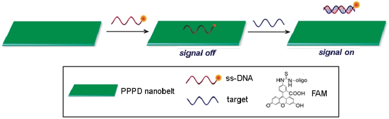
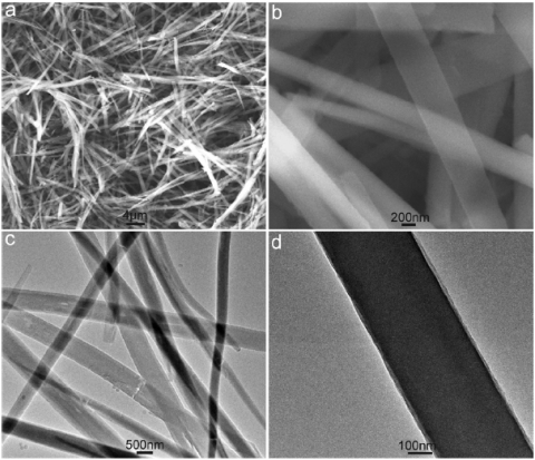
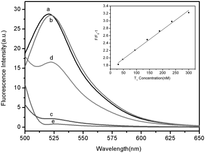
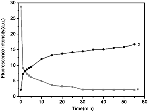
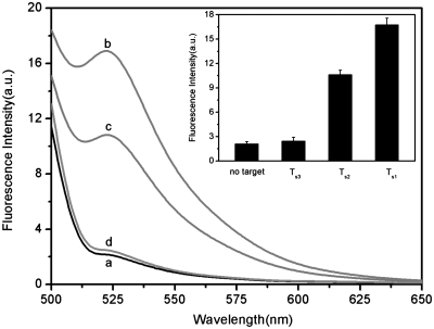
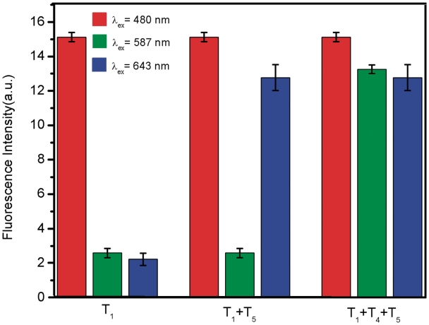
In this article, we report on the facile and rapid synthesis of conjugation polymer poly(p-phenylenediamine) nanobelts (PNs) via room temperature chemical oxidation polymerization of p-phenylenediamine monomers by ammonium persulfate in aqueous medium. We further demonstrate the proof-of-concept that PNs can be used as an effective fluorescent sensing platform for nucleic acid detection for the first time. The general concept used in this approach lies in the facts that the adsorption of the fluorescently labeled single-stranded DNA probe by PN leads to substantial fluorescence quenching, followed by specific hybridization with the complementary region of the target DNA sequence. This results in desorption of the hybridized complex from PN surface and subsequent recovery of fluorescence. We also show that the sensing platform described herein can be used for multiplexing detection of nucleic acid sequences.


Figures

Scheme 1.
Scheme 1.
A schematic diagram (not to scale) to illustrate the fluorescence-enhanced nucleic acid detection using PPPD nanobelt as a sensing platform.
Figure 1.
Figure 1.
Low magnification SEM (a) and TEM (c) images of the products thus formed. (b and d) Indicates the corresponding high magnification images.
Figure 2.
Figure 2.
Fluorescence emission spectra of PHIV (50 nM) at different conditions: (a) PHIV; (b) PHIV + 300 nM T1; (c) PHIV + PN; (d) PHIV + PN + 300 nM T1. Curve e is the emission spectra of PN. Inset: linear relationship between F/F0−1 (where F0 and F are the fluorescence intensity without and with the presence of T1, respectively) and T1 concentration ranging from 30 to 300 nm. Excitation was at 480 nm, and the emission was monitored at 522 nm. All measurements were done in Tris–HCl buffer in the presence of 5 mM Mg2+ (pH: 7.4).
Figure 3.
Figure 3.
(a) Fluorescence quenching of PHIV (50 nM) by PN and (b) fluorescence recovery of PHIV–PN by T1 (300 nM) as a function of incubation time. Excitation was at 480 nm, and the emission was monitored at 522 nm. All measurements were done in Tris–HCl buffer in the presence of 5 mM Mg2+ (pH: 7.4).
Figure 4.
Figure 4.
(a) Fluorescence emission spectra of PHIV (50 nM) at different conditions: (a) PHIV–PN complex; (b) PHIV–PN complex + 300 nM T1; (c) PHIV–PN complex + 300 nM T2; (d) PHIV–PN complex + 300 nM T3. Inset: fluorescence intensity histograms with error bar. (b) Fluorescence signal enhancement of PHIV–PN complex upon incubation with T1 and T2 at 25 and 50°C, respectively. Excitation was at 480 nm, and the emission was monitored at 522 nm. All measurements were done in Tris–HCl buffer in the presence of 5 mM Mg2+ (pH: 7.4).
Figure 5.
Figure 5.
(a) Fluorescence emission spectra of Ps (50 nM) at different conditions: (a) Ps–PN complex; (b) Ps–PN complex + 300 nM Ts1; (c) Ps–PN complex + 300 nM Ts2; (d) Ps–PN complex + 300 nM Ts3. Inset: fluorescence intensity histograms with error bar.
Figure 6.
Figure 6.
Fluorescence intensity histograms of the probe mixture toward different target combinations in the presence of PN under excitation/emission wavelengths of 480/522, 587/606 and 643/665 nm/nm. All measurements were done in Tris–HCl buffer in the presence of 5 mM Mg2+ (pH: 7.4).

References

    1. Gresham D, Ruderfer DM, Pratt SC, Schacherer J, Dunham MJ, Botstein D, Kruglyak L. Genome-wide detection of polymorphisms at nucleotide resolution with a single DNA microarray. Science. 2006;311:1932–1936. - PubMed
    1. Sassolas A, Leca-Bouvier BD, Blum LJ. DNA biosensors and microarrays. Chem. Rev. 2008;108:109–139. - PubMed
    1. Didenko VV, editor. Fluorescent Energy Transfer Nucleic Acid Probes: designs and Protocols. Totowa, NJ: Human Press; 2006.
    1. Holland PM, Abramson RD, Watson R, Gelfand DH. Detection of specific polymerase chain reaction product by utilizing the 5′→3′ exonuclease activity of Thermus aquaticus DNA polymerase. Proc. Natl Acad. Sci. USA. 1991;88:7276–7280. - PMC - PubMed
    1. Tyagi S, Kramer FR. Molecular beacons: probes that fluoresce upon hybridization. Nat. Biotechnol. 1996;14:303–308. - PubMed

Publication types

MeSH terms