. 2011 Apr;7(4):386-400.
doi: 10.4161/auto.7.4.14391. Epub 2011 Apr 1.

The Atg6/Vps30/Beclin 1 ortholog BEC-1 mediates endocytic retrograde transport in addition to autophagy in C. elegans

Affiliations

The Atg6/Vps30/Beclin 1 ortholog BEC-1 mediates endocytic retrograde transport in addition to autophagy in C. elegans

Alexander Ruck et al. Autophagy. 2011 Apr.

Abstract

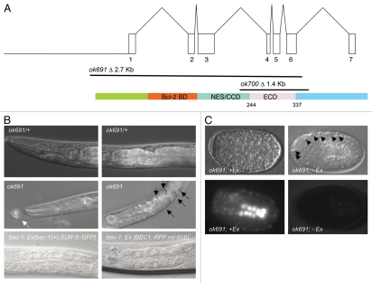
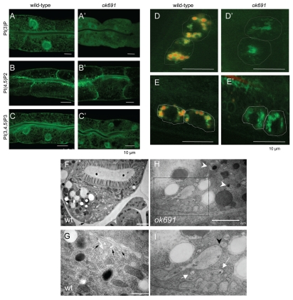
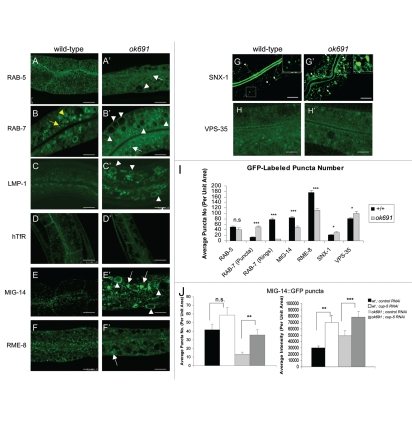
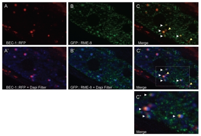
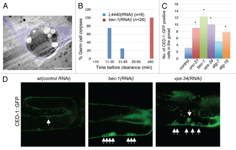
Autophagy and endocytosis are dynamic and tightly regulated processes that contribute to many fundamental aspects of biology including survival, longevity, and development. However, the molecular links between autophagy and endocytosis are not well understood. Here, we report that BEC-1, the C. elegans ortholog of Atg6/Vps30/Beclin1, a key regulator of the autophagic machinery, also contributes to endosome function. In particular we identify a defect in retrograde transport from endosomes to the Golgi in bec-1 mutants. MIG-14/Wntless is normally recycled from endosomes to the Golgi through the action of the retromer complex and its associated factor RME-8. Lack of retromer or RME-8 activity results in the aberrant transport of MIG-14/Wntless to the lysosome where it is degraded. Similarly, we find that lack of bec-1 also results in mislocalization and degradation of MIG-14::GFP, reduced levels of RME-8 on endosomal membranes, and the accumulation of morphologically abnormal endosomes. A similar phenotype was observed in animals treated with dsRNA against vps-34. We further identify a requirement for BEC-1 in the clearance of apoptotic corpses in the hermaphrodite gonad, suggesting a role for BEC-1 in phagosome maturation, a process that appears to depend upon retrograde transport. In addition, autophagy genes may also be required for cell corpse clearance, as we find that RNAi against atg-18 or unc-51 also results in a lack of cell corpse clearance.


Figures

Figure 1
Figure 1
bec-1 gene, protein, mutations and phenotypes. (A) At the top, the genomic structure of the bec-1 gene encompassing 7 exons, and encoding a 375 amino acid protein, shown below. In the middle, two deletions of bec-1 as provided by the C. elegans Gene Knockout Consortium. ok691 deletes most of the bec-1 open reading frame including the start ATG and exons 1–6, thus it is a molecular null. ok700 deletes exons 4–6 of bec-1 and renders the mRNA out of frame for exon 7. A cDNA was sequenced after RNA was extracted from an ok700 lysate, which shows that there is bec-1 mRNA transcribed in ok700 mutants that lacks the evolutionarily conserved domain of BEC-1 required for binding VPS-34. (B) Both deletions display an adult lethal phenotype with a striking accumulation of vacuoles (marked by black arrows in (B), middle, right panel). ok691 mutants (B, middle panel) segregating from a heterozygous parent (B, top panel). bec-1 homozygous mutant, either ok691 or ok700, appear superficially wild type during early larval stages but become increasingly uncoordinated during later larval stages and display molting problems (white arrow in (B), middle, left panel). Rescue of the bec-1 phenotypes including uncoordination, the accumulation of vacuoles and lethality was observed with a transgene containing the bec-1 wild-type genomic sequences and a ubiquitously expressed SUR-5::GFP marker (B, bottom, left panel) or a BEC-1::RFP construct (B, bottom, right panel). (C) Animals that carry the rescuing bec-1 genomic extrachromosomal array (Ex[bec-1(+), SUR-5::GFP]) (left bottom panel) are rescued for the bec-1 phenotype. Animals that have lost the array early in development are completely devoid of the GFP marker expression (C, right bottom panel) and lack the maternal and zygotic bec-1 expression. A hatched line delineates the periphery of the embryo (C, right bottom panel). These animals die during embryogenesis with an increase in the number of visible apoptotic cells (marked by black arrow heads, (C) top right panel).
Figure 2
Figure 2
bec-1 mutants display defects in endocytosis. The detection of the phosphoinositides PI3P, PIP2 and PIP3 in the intestines of wild-type (A–C), bec-1(ok691) mutants (A'–C'), animals is shown. Scale bars indicate 10 µm. In wild-type animals (A), PI3P is enriched in the apical membrane with weak basolateral labeling. In bec-1(ok691) (A') mutants very low and diffused expression of PI3P was observed. PIP2 expression is not affected in bec-1(ok691) mutants (B') when compared to wild-type animals (B). The PIP3 reporter labels apical membrane and strong basolateral expression in wild-type animals (C). The localization of PIP3 in bec-1(ok691) animals (C') was similar to that of wild-type animals (C). Micrographs of LMP-1::GFP positive lysosomes in coelomocytes 24 h after the injection of Texas Red BSA (a fluid phase marker for endocytosis) into the body cavity (pseudocoelom). A hatched line demarcates the periphery of the coeolomocytes. The red dye accumulates in lysosomes (LMP-1 positive) localized in the coelomocytes of the wild-type animals (D), but fails to accumulate in the lysosomes (LMP-1 positive) of bec-1(ok691) mutants (D'). Micrographs of LMP-1::GFP positive lysosomes in coelomocytes after 1 h of soaking in Texas Red BSA (a fluid phase marker for endocytosis). The red dye accumulates in lysosomes (green) localized in the coelomocytes of the wild-type animals (E), but fails to accumulate in the lysosomes (green) of bec-1 mutants (E'). Electron microscopy of an N2 wild-type (F and G) and bec-1(ok691) mutant young adults (H and I). (F) Low power electron micrograph of wild-type adult intestine in cross-section, fixed by high pressure freezing. Most of the basal membrane shows few signs of endocytosis, although there is some membrane infolding (black arrow in F) and a few small vesicles, particularly near the lateral membrane border between two intestinal cells on the extreme left edge of the tissue. Higher power view of adult wild-type intestine shows a few examples of membrane infolding (black arrows in G) representing a local zone where endocytosis is active. It is notable that the infolded membranes are rather large, and there are almost no vesicles nearby in the cytoplasm. In contrast, chains of vesicles (white arrows in I) and extensive infolded membranes are seen along the basal pole in the bec-1 mutant (H and I). Thus endocytotic events must be cleared relatively quickly in the wild-type intestine. In bec-1 mutant intestines, defective enlarged endosomes (marked by white arrow head in H) are seen that correspond to abnormal vacuoles of different sizes and contain either membranes or condensed material. (I) is a close up of the region marked in (H) and it shows enlarged vacuoles and a high number of early endosomes that appear to be fusing in some cases (black arrowhead in I). This phenotype is never seen in the intestines of wild-type animals (F or G).
Figure 3
Figure 3
bec-1 mutants display a defect in retrograde transport. Confocal images in a wild-type background are shown for GFP::RAB-5 (A), GFP::RAB-7 (B), LMP-1::GFP (C), GFP::hTfR (D), MIG-14::GFP (E), GFP::RME-8 (F), GFP::SNX-1 (G), GFP::VPS-35 (H). Confocal images of bec-1(ok691) mutants are shown for GFP::RAB-5 (A'), GFP::RAB-7 (B'), LMP-1::GFP (C'), GFP::hTfR (D'), MIG-14::GFP (E'), RME-8::GFP (F'), GFP::SNX-1 (G'), GFP::VPS-35 (H'). (B) GFP::RAB-7 positive puncta (marked by yellow arrow) labels maturing endosomes and late endosomes (marked by yellow arrowhead). bec-1(ok691) mutants accumulate maturing endosomes marked with GFP::RAB-7, and display a decrease in late endosomes marked with GFP::RAB-7 (compare B and B'; See Fig. 2I). White arrowheads indicate enlarged intestinal endosomes (abnormal vacuoles) labeled by GFP::RAB-5 and GFP::RAB-7. In bec-1(ok691) animals, LMP-1::GFP also accumulates in the membrane of the enlarged endosomes (C'; marked by white arrowheads). In wild-type animals (D), the basolateral plasma membrane and basolateral endocytic compartments are labeled by hTfR::GFP (human transferrin receptor). The accumulation of hTfR::GFP is not affected in bec-1(ok691) mutants (D'), and the recycling endosome cargo hTfR::GFP does not accumulate in the enlarged endosomes (abnormal vacuoles) of bec-1(ok691) mutants. Confocal images of the worm intestine expressing the GFP-tagged endocytic transmembrane cargo marker MIG-14::GFP in wild-type (E), and bec-1(ok691) (E') animals. The retromer-dependent cargo protein Wntless MIG-14::GFP normally localizes basolaterally in the intestine and colocalizes with the early endosomal marker RAB-5. In bec-1 mutants, MIG-14::GFP transmembrane cargo protein accumulates in the enlarged endosomes (abnormal vacuoles; E'). We observed a decrease in the number of MIG-14::GFP positive puncta (I). The retromer subunit RME-8::GFP (F'), GFP::SNX-1 (G') and GFP::VPS-35 (H'), do not accumulate in the enlarged intestinal endosomes (abnormal vacuoles). A decrease in the number of GFP::RME-8 positive puncta is observed in bec-1(ok691) mutants (F'), and its intensity is markedly decreased. For all images, scale bars represent 10 µm. Quantification of endosome number as visualized by the positive labeling with endocytic markers is shown in (I). In the quantification of endosomal compartments, we saw no significant difference in RAB-5 early endosomes, we observed an increase in RAB-7 maturing endosomes, and a dramatic decrease in RAB-7-positive late endosomes. In addition, we observed a decrease in MIG-14 as well as RME-8 labeled compartments, and a slight increase in SNX-1 as well as in VPS-35 labeled compartments. Error bars represent standard deviation from the mean (n = 30 each, 10 animals of each genotype were sampled in three different regions of the intestine). In (J and K), we analyzed whether lysosomal degradation of GFP::MIG-14 occurs in bec-1 mutant animals. RNAi mediated depletion of lysosome biogenesis protein CUP-5/mucolipin1 increased the number and the intensity of MIG-14::GFP positive puncta (J and Fig. S5). After cup-5 RNAi, we saw a 4X increase in the number of MIG-14 positive puncta in bec-1 mutants, when compared to control RNAi, indicating that there is lysosomal processing of the MIG-14 cargo in bec-1 mutants (J, left panel). Similarly, we observed a significant increase in the intensity of MIG-14::GFP in bec-1 mutants after cup-5 RNAi (J, right panel). Asterisks indicate a significant difference in the one-tailed Student's t-test (*p < 0.05, **p < 0.005, ***p < 0.0005, n.s.: not significant).
Figure 4
Figure 4
BEC-1 colocalizes with RME-8. Representative images of intestinally expressed RFP-tagged BEC-1 (red, A, A') and GFP-tagged RME-8 (green, B, B') in wild-type intact living animals. Autofluorescent lysosome-like organelles are shown in blue (+ DAPI filter, D). White arrowheads indicate colocalization of the BEC-1::RFP and RME-8::GFP signals (C), observed as the yellow fluorescence. The blue autofluorescence is also shown with BEC-1::RFP (A') and with GFP::RME-8 (B') and in the merger of both in (C'). The colocalized BEC-1::RFP and GFP::RME-8 signals are found adjacent to the autofluorescent lysosomes which can be visualized with the DAPI filter (C'). A close-up of (C') is shown in (C”). Arrowheads in yellow show BEC-1::RFP that is not associated with RME-8::GFP. Magnification is 630x (A–C').
Figure 5
Figure 5
bec-1 mutants display lack of cell corpse clearance. (A) Micrograph of transmission electron microscopy (TEM) where poor degradation of apoptotic germ cell corpses is observed in bec-1 mutants. This TEM image was taken in a cross-section through the midbody. A rounded dying germ cell is shown with very dark cytoplasm and several large round vacuoles near the nucleus, which contains clumped chromatin. The dying cell is completely wrapped by the somatic sheath cell (tinted in purple) in an early phase of apoptosis. The sheath cell also wraps the normal germ-line (bottom two cells in the panel), separating this mesodermal tissue from the intestine (top cell in the panel). (B) Histogram indicating the distribution of the duration of germ cell corpses before they are completely degraded. The y-axis indicates the percentage of germ cell corpses that lasted for the period of time indicated in the x-axis before clearance. n is the number of cell corpses analyzed. (C) Representative images of CED-1::GFP positive labeled cell corpses in wild-type animals after treatment with control RNAi and RNAi against bec-1 and vps-34. (D) Quantification of germ cell corpses in wild-type animals after treatment with control RNAi, and RNAi against unc-51/Atg1, bec-1, vps-34, atg-7 and atg-18. Data derived from observing adults, 36 hours post larval L4 stage. Data were compared by unpaired t tests; 30 animals were analyzed for each experiment. Asterisks indicate a significant difference as a result from an analysis of variance (ANOVA). This analysis indicated that there are significant differences between all treatments and control, except for atg-7 RNAi (p < 0.001). Threshold for significance (alpha) in the t-tests was p < 0.01 using a “Bonferroni correction” for multiple corrections.

References

    1. Mizushima N, Levine B, Cuervo AM, Klionsky DJ. Autophagy fights disease through cellular self-digestion. Nature. 2008;451:1069–1075. - PMC - PubMed
    1. Melendez A, Neufeld TP. The cell biology of autophagy in metazoans: a developing story. Development. 2008;135:2347–2360. - PMC - PubMed
    1. Suzuki K, Kubota Y, Sekito T, Ohsumi Y. Hierarchy of Atg proteins in pre-autophagosomal structure organization. Genes Cells. 2007;12:209–218. - PubMed
    1. Klionsky DJ, Cregg JM, Dunn W, Jr, Emr SD, Sakai Y, Sandoval IV, et al. A unified nomenclature for yeast autophagy-related genes. Dev Cell. 2003;5:539–545. - PubMed
    1. Suzuki K, Kirisako T, Kamada Y, Mizushima N, Noda T, Ohsumi Y. The pre-autophagosomal structure organized by concerted functions of APG genes is essential for autophagosome formation. EMBO J. 2001;20:5971–5981. - PMC - PubMed

Publication types

Substances

LinkOut - more resources