Review
. 2011 May;37(3):377-86.
doi: 10.1016/j.burns.2010.11.012. Epub 2010 Dec 23.

Noninvasive assessment of burn wound severity using optical technology: a review of current and future modalities

Affiliations
Review

Noninvasive assessment of burn wound severity using optical technology: a review of current and future modalities

Meghann Kaiser et al. Burns. 2011 May.

Abstract

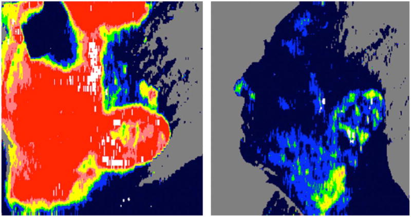
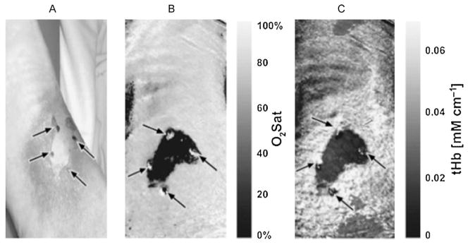
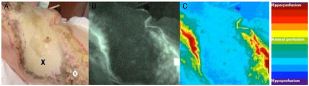
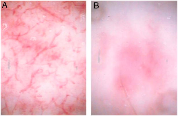
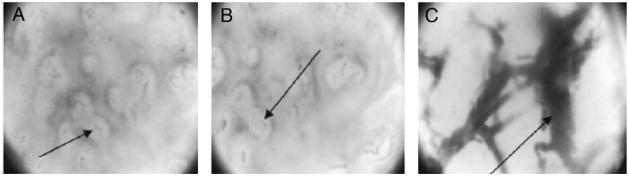
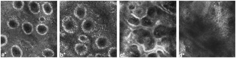
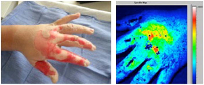
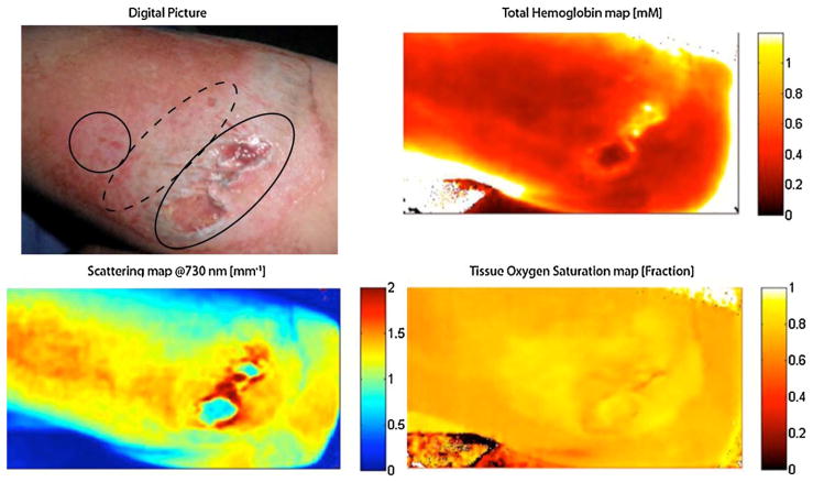
Clinical examination alone is not always sufficient to determine which burn wounds will heal spontaneously and which will require surgical intervention for optimal outcome. We present a review of optical modalities currently in clinical use and under development to assist burn surgeons in assessing burn wound severity, including conventional histology/light microscopy, laser Doppler imaging, indocyanine green videoangiography, near-infrared spectroscopy and spectral imaging, in vivo capillary microscopy, orthogonal polarization spectral imaging, reflectance-mode confocal microscopy, laser speckle imaging, spatial frequency domain imaging, photoacoustic microscopy, and polarization-sensitive optical coherence tomography.


Conflict of interest statement

Conflict of interest

Dr. Anthony Durkin has a financial interest in Modulated Imaging Inc., a company with interests related to spatial field domain imaging (SFDI). Dr. Durkin is a cofounder of the company and owns equity interests in Modulated Imaging.

Figures

Fig. 1
Fig. 1
On the left, LDI-generated blood flow map of a superficial partial thickness burn on a patient’s face. High blood flow is manifest as a predominantly bright red region. On the right, the same area on the same patient minutes later, showing significantly diminished perfusion, as suggested by the now dark blue facial area. Shortly after the second scan, the patient experienced a near-syncopal episode. (For interpretation of the references to color in this figure legend, the reader is referred to the web version of the article.)
Fig. 2
Fig. 2
Near-infrared spectral imaging. (A) Digital photograph of burn wound to the leg with central area of full thickness. (B) Map of tissue oxygen saturation with darker areas representing less oxygenation, corresponding to full thickness injury. (C) Map of total hemoglobin concentration with darker areas representing less hemoglobin. (Reprinted from Ref. [40]. Copyright 2007 by John Wiley and Sons. Reprinted with permission.)
Fig. 3
Fig. 3
Indocyanine green video angiography. (A) Digital photograph of a burn to the chest with central area of full thickness (x) and peripheral areas of partial thickness (o). (B) Raw angiography image with brighter areas corresponding to regions of increased perfusion. (C) Computer generated image quantifying fluorescence with areas of greater perfusion represented by red and less perfusion by blue. (Reprinted from Ref. [29]. Copyright 2003 by Elsevier. Reprinted with permission.) (For interpretation of the references to color in this figure legend, the reader is referred to the web version of the article.)
Fig. 4
Fig. 4
In vivo capillary microscopy. (A) Superficial burn with preserved dermal capillary plexus visible. (B) Deep partial thickness burn with absence of the capillary plexus apparent. (Reprinted from Ref. [22]. Copyright 2007 by Elsevier. Reprinted with permission.)
Fig. 5
Fig. 5
Orthogonal polarization spectral imaging. (A) Normal skin, in which the dermal capillary plexus is apparent as faint gray loops. (B) Superficial burn, appearing very similar to normal skin. (C) Deep burn, in which the destruction of the overlying capillary plexus allows for visualization of the larger thrombosed dermal vessels beneath. (Reprinted from Ref. [43]. Copyright 2005 by Elsevier. Reprinted with permission.)
Fig. 6
Fig. 6
Reflectance confocal microscopy of a human subject at 24 h post-injury showing (a) normal skin, with dermal capillaries manifested as black punctuate areas within dermal papillae (white rings), (b) superficial burn, with apparent enlargement of the dermal capillaries secondary to edema, (c) superficial partial-thickness burn, showing incomplete destruction of the dermal papillae, and (d) deep partial-thickness burn, showing complete destruction of the papillae. (Reprinted from Ref. [47]. Copyright 2009 by Elsevier. Reprinted with permission.)
Fig. 7
Fig. 7
On the left, a digital photograph of a hot oil burn to the hand with areas of deep (white) and partial (pink) thickness. On the right, the laser speckle image (LSI) of the same burn. Areas of high perfusion are represented by brighter colors (red, yellow, green, in order of decreasing perfusion) and lower perfusion is represented by blue. Deep thickness regions on the proximal second, third, and fourth digits correspond to light blue areas on the speckle image, whereas the dorsum of the hand overlying the metacarpals is partial thickness on clinical exam green/yellow/red on the speckle image. Uninjured, non-inflamed skin is blue. (Patient permission was obtained to publish photograph.) (For interpretation of the references to color in this figure legend, the reader is referred to the web version of the article.)
Fig. 8
Fig. 8
In the upper left corner, a digital photograph of mechanical burn (road rash) to the forearm with areas of full (oval), deep partial (dashed oval), and superficial partial (circle) thickness injury. Regional maps of hemoglobin, oxygen saturation, and light scattering can differentiate between different depths within the wound as shown (patient permission was obtained to publish photograph).

References

    1. Janzekovic Z. A new concept in the early excision and immediate grafting of burns. J Trauma. 1970;10(12):1103–8. - PubMed
    1. Cramer LM, McCormack RM, Carroll DB. Progressive partial excision and early grafting in lethal burns. Plast Reconstr Surg Transplant Bull. 1962;30:595–9. - PubMed
    1. Meeker IA, Snyder WH. Dermatome debridement and early grafting of extensive third degree burns in children. Surg Gynelcol Obstet. 1956;103(5):527–34. - PubMed
    1. Saffle JR. Practice guidelines for burn care. J Burn Care Rehabil. 2001;22(Suppl 1)
    1. Orgill DP. Excision and skin grafting of thermal burns. N Engl J Med. 2009;360(9):893–901. - PubMed

Publication types