Review
. 2012 Jan;1822(1):21-33.
doi: 10.1016/j.bbadis.2010.12.014. Epub 2010 Dec 23.

Mast cells and inflammation

Affiliations
Review

Mast cells and inflammation

Theoharis C Theoharides et al. Biochim Biophys Acta. 2012 Jan.

Abstract

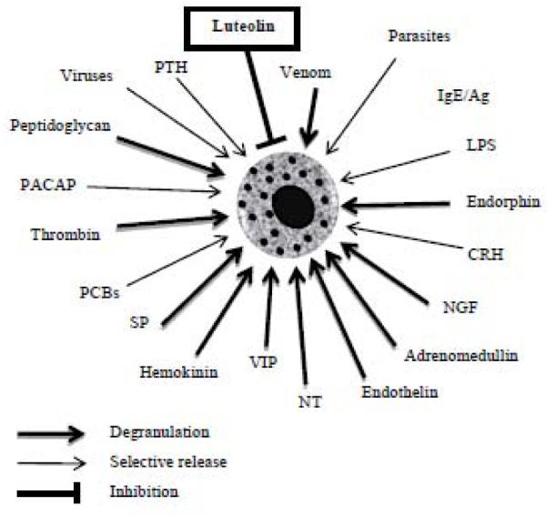
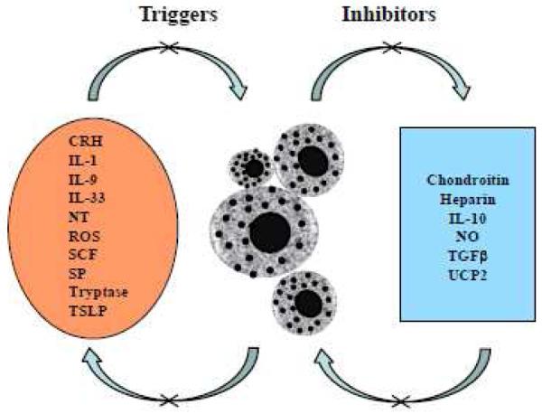
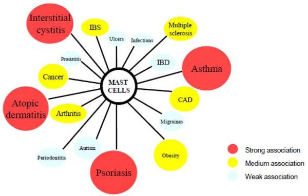
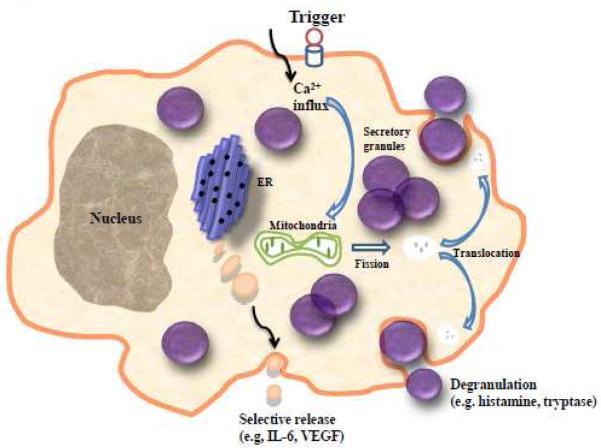
Mast cells are well known for their role in allergic and anaphylactic reactions, as well as their involvement in acquired and innate immunity. Increasing evidence now implicates mast cells in inflammatory diseases where they are activated by non-allergic triggers, such as neuropeptides and cytokines, often exerting synergistic effects as in the case of IL-33 and neurotensin. Mast cells can also release pro-inflammatory mediators selectively without degranulation. In particular, IL-1 induces selective release of IL-6, while corticotropin-releasing hormone secreted under stress induces the release of vascular endothelial growth factor. Many inflammatory diseases involve mast cells in cross-talk with T cells, such as atopic dermatitis, psoriasis and multiple sclerosis, which all worsen by stress. How mast cell differential responses are regulated is still unresolved. Preliminary evidence suggests that mitochondrial function and dynamics control mast cell degranulation, but not selective release. Recent findings also indicate that mast cells have immunomodulatory properties. Understanding selective release of mediators could explain how mast cells participate in numerous diverse biologic processes, and how they exert both immunostimulatory and immunosuppressive actions. Unraveling selective mast cell secretion could also help develop unique mast cell inhibitors with novel therapeutic applications. This article is part of a Special Issue entitled: Mast cells in inflammation.


Figures

Figure 1
Figure 1
Schematic representation of physiological and environmental mast cell triggers, and the inhibitory effect of certain flavonoids, such as luteolin. Many of these triggers stimulate selective release of mediators such as IL-6, TNF or VEGF without degranulation. CRH, corticotropin releasing hormone; LPS, lipopolysaccharide; NT, neurotensin; PACAP, pituitary adenylate cyclase activating polypeptide; PCBs, polychlorinated biphenols; PTH, parathyroid hormone; SP, substance P; VIP, vasoactive intestinal peptide.
Figure 2
Figure 2
Schematic representation of mast cell autocrine triggers and modulators. Numerous molecules secreted by mast cells can have autocrine actions, either activating or inhibiting mast cells. CRH, corticotropin-releasing hormone; IL, interleukin; NT, neurotensin; NO, nitic oxide; ROS, reactive oxygen species; SCF, stem cell factor; SP, substance P; TGFβ, transforming growth factor β; TSLP, thymic stromal lymphopoietin; UCP2, uncoupling protein 2.
Figure 3
Figure 3
Mast cell involvement in inflammatory diseases. Increasing evidence indicates that mast cells are involved in many diseases. Colors indicate the strength of the association (red = strongest, white = weakest). CAD, coronary artery disease; IBD, inflammatory bowel disease; IBS, irritable bowel syndrome.
Figure 4
Figure 4
Schematic representation showing mast cell degranulation as compared to selective mediator release. During selective release, vesicles much smaller than secretory granules transport mediators to the cell surface for exocytosis. ER, endoplasmic reticulum; VEGF, vascular endothelial growth factor.
Figure 5
Figure 5
Two human cultured LAD2 mast cells, showing distribution of mitochondria stained with MitoTracker and photographed using Confocal microscopy; (A) control in which mitochondria form a “net” around the nucleus and (B) after stimulation with SP (2 M for 30 min at 37°C) in which mitochondria are distributed throughout the cell. (Magnification: x 1000). Arrows point to the areas with the highest concentration of MitoTracker (yellow color); thus the highest aggregation of mitochondria.

References

    1. Rodewald HR, Dessing M, Dvorak AM, Galli SJ. Identification of a committed precursor for the mast cell lineage. Science. 1996;271:818–822. - PubMed
    1. Chen CC, Grimbaldeston MA, Tsai M, Weissman IL, Galli SJ. Identification of mast cell progenitors in adult mice. Proc Natl Acad Sci U.S.A. 2005;102:11408–11413. - PMC - PubMed
    1. Kitamura Y, Ito A. Mast cell-committed progenitors. Proc Natl Acad Sci U.S.A. 2005;102:11129–11130. - PMC - PubMed
    1. Hundley TR, Gilfillan AM, Tkaczyk C, Andrade MV, Metcalfe DD, Beaven MA. Kit and FcepsilonRI mediate unique and convergent signals for release of inflammatory mediators from human mast cells. Blood. 2004;104:2410–2417. - PubMed
    1. Blank U, Rivera J. The ins and outs of IgE-dependent mast-cell exocytosis. Trends Immunol. 2004;25:266–273. - PubMed

Publication types

Substances

LinkOut - more resources