Review
. 2011 Jan 4;57(1):9-17.
doi: 10.1016/j.jacc.2010.08.623.

The aging heart and post-infarction left ventricular remodeling

Affiliations
Review

The aging heart and post-infarction left ventricular remodeling

Henry Shih et al. J Am Coll Cardiol. .

Abstract

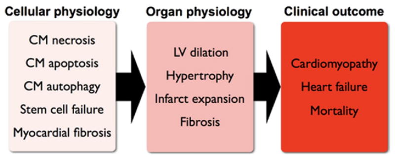
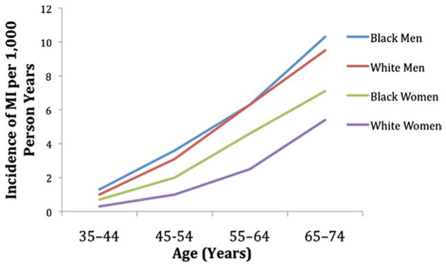
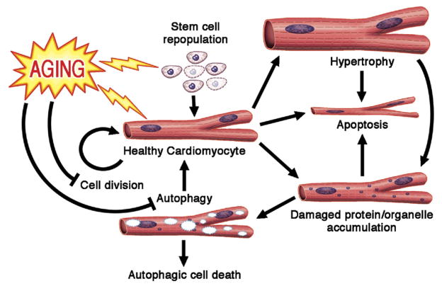
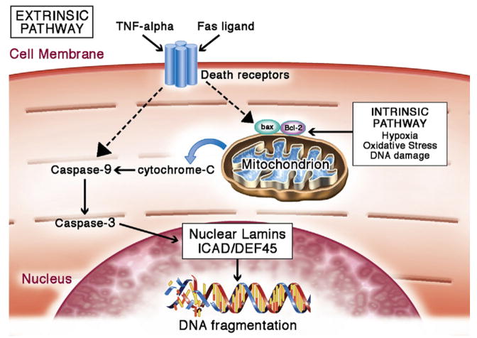
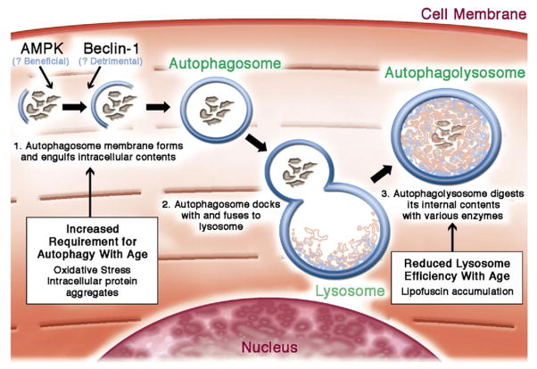
Aging is a risk factor for heart failure, which is a leading cause of death world-wide. Elderly patients are more likely than young patients to experience a myocardial infarction (MI) and are more likely to develop heart failure following MI. The poor clinical outcome of aging in cardiovascular disease is recapitulated on the cellular level. Increase in stress exposure and shifts in signaling pathways with age change the biology of cardiomyocytes. The progressive accumulation of metabolic waste and damaged organelles in cardiomyocytes blocks the intracellular recycling process of autophagy and increases the cell's propensity toward apoptosis. Additionally, the decreased cardiomyocyte renewal capacity in the elderly, due to reduction in cellular division and impaired stem cell function, leads to further cardiac dysfunction and maladaptive responses to disease or stress. We review the cellular and molecular aspects of post-infarction remodeling in the aged heart, and relate them to the clinical problem of post-infarction remodeling in elderly patients.


Figures

Figure 1
Figure 1. Post-MI Remodeling of the Heart at Cellular and Organ Levels
CM = cardiomyocyte; LV = left ventricular.
Figure 2
Figure 2. Increased Rates of MI With Age
Incidence of myocardial infarction (MI) increases with age, and is higher in men than in women. Data derived from ARIC (Atherosclerosis Risk in Communities) surveillance (5).
Figure 3
Figure 3. Aging of Cardiomyocytes
Aging is characterized with decreased cardiomyocyte renewal, possibly through the impairment of both the stem cell differentiation into cardiomyoctyes and cardiomyocyte division. Additionally, changes in cardiomyocyte microenvironment and signaling pathways increase the cell’s propensity toward apoptosis. Progressive accumulation of damaged protein and organelles in cardiomyoctyes blocks autophagy mechinary. Failure to clear out the intracellular waste can induce apoptosis of the cell. Compensatory neurohormonal signals induce the remaining cardiomyocytes to undergo hypertrophy and increase metabolic demand, thereby exposing the cellular contents to oxidative damage. Regular arrows = promotion/induction; blocked arrows = inhibition; lightning = detrimental effects of aging. Figure illustration by Craig Skaggs.
Figure 4
Figure 4. Cardiomyocyte Apoptosis
Apoptosis can be mediated by the intrinsic or extrinsic pathways. Many factors associated with these pathways are increased with aging, resulting in a predisposition toward apoptosis in the aging cardiomyocyte. Intrinsically, apoptosis is activated in the event of hypoxia, oxidative stress, DNA damage, acidosis, or growth factor deprivation (36,37). These stressors activate cell death signaling chiefly through Bcl-2 family–regulated increase of mitochondrial outer membrane permeabilization (MOMP). Cytochrome C from mitochondria diffuses into the cytoplasm following the increase in mitochondrial membrane permeability. In the cytosol, cytochrome C activates a downstream cascade to initiate caspase activities (38,39). Active caspases target nuclear lamins, nuclear inhibitor of caspase-activated deoxyribonuclease/DNA fragmentation factor-45 (ICAD/DEF45), and various other cytoplasmic proteins. Cleavage of nuclear lamins breaks down the nuclear structure (40). Cleavage of ICAD/DEF45, a DNase inhibitor, disinhibits DNase, thereby allowing it to break down DNA (41). Extrinsic apoptosis can be activated by Fas ligand or tumor necrosis factor (TNF)-alpha. Binding of TNF-alpha or Fas ligand to their respective receptors activates TNF receptor–associated death domain (TRADD) and Fas-associated death domain (FADD) proteins (42,43). These proteins act on regulators of mitochondria to change the permeability of the mitochondrial outer membrane, and this change converges with intrinsic apoptosis activation on the caspase cascade. Figure illustration by Craig Skaggs.
Figure 5
Figure 5. Cardiomyocyte Autophagy
Autophagy may be triggered by the accumulation of intracellular protein aggregation (48), nutrient deprivation (49), or various intracellular signals. The process initiates with a highly regulated activation step that forms a double membrane autophagosome, which engulfs damaged proteins and organelles. Autophagosomes then fuse with the lysosome, which contain proteases to digest the engulfed content. Aging increases the cardiomyocyte’s need for autopahgy to maintain intracellular homeostasis, but simultaneously reduces the activity of lysosomes and thereby inhibits autophagic flux. Figure illustration by Craig Skaggs.

References

    1. National Institutes of Health. National Heart Lung and Blood Institute Factbook Fiscal Year 2008. [Accessed June 1, 2010]. Available at: http://www.nhlbi.nih.gov/about/factbook/FactBookFinal.pdf.
    1. Lloyd-Jones D, Adams RJ, Brown TM, et al. Heart disease and stroke statistics—2010 update: a report from the American Heart Association. Circulation. 2010;121:e46–215. - PubMed
    1. Yousef ZR, Redwood SR, Marber MS. Postinfarction left ventricular remodelling: where are the theories and trials leading us? Heart. 2000;83:76–80. - PMC - PubMed
    1. Administration on Aging. A Profile of Older Americans. Department of Health and Human Services; 2008. [Accessed June 1, 2010]. Available at: http://www.csrees.usda.gov/nea/economics/pdfs/profile_older_american_200....
    1. National Heart, Lung, and Blood Institute. Incidence and Prevalence: 2006 Chart Book on Cardiovascular and Lung Diseases. Bethesda, MD: National Institutes of Health; 2006.

Publication types