. 2011 Feb;46(2):535-47.
doi: 10.1016/j.mcn.2010.12.010. Epub 2010 Dec 24.

NFAT regulates pre-synaptic development and activity-dependent plasticity in Drosophila

Affiliations

NFAT regulates pre-synaptic development and activity-dependent plasticity in Drosophila

Amanda Freeman et al. Mol Cell Neurosci. 2011 Feb.

Abstract

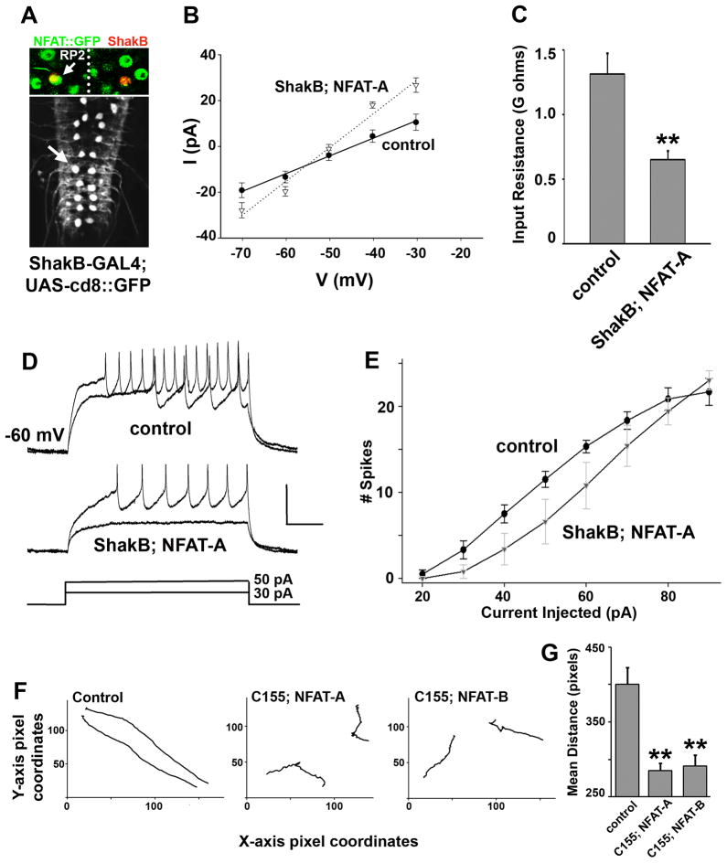
The calcium-regulated transcription factor NFAT is emerging as a key regulator of neuronal development and plasticity but precise cellular consequences of NFAT function remain poorly understood. Here, we report that the single Drosophila NFAT homolog is widely expressed in the nervous system including motor neurons and unexpectedly controls neural excitability. Likely due to this effect on excitability, NFAT regulates overall larval locomotion and both chronic and acute forms of activity-dependent plasticity at the larval glutamatergic neuro-muscular synapse. Specifically, NFAT-dependent synaptic phenotypes include changes in the number of pre-synaptic boutons, stable modifications in synaptic microtubule architecture and pre-synaptic transmitter release, while no evidence is found for synaptic retraction or alterations in the level of the synaptic cell adhesion molecule FasII. We propose that NFAT regulates pre-synaptic development and constrains long-term plasticity by dampening neuronal excitability.


Figures

Figure 1
Figure 1. Drosophila NFAT is expressed in the nervous system
A) Schematic of the NFAT gene showing exon-intron boundaries and two NFAT transcripts, NFAT-A and NFAT-B (specific riboprobes to these transcripts are designated RP-A and RP-B). Also shown are insertion sites for the two EP elements (19579 and 1508) that predictably drive expression of the NFAT-A or NFAT-B isoforms respectively, the site of insertion of the GFP exon trap element, and the region targeted by the NFAT-RNAi construct. The conserved Rel-Homology Domain (RHD) spans exons 5 and 6 and the NFAT deletion ΔAB used in this study eliminates shared exons 2 and 3. B) Western blot of brain protein extracts probed with antibodies raised against the C-terminal of NFAT. A band of the predicted molecular weight (~150 KD) is recognized in wild type brains, is absent in the deletion mutant heterozygous over a non-complementing deficiency and is present in higher level when either NFAT-A or NFAT-B is overexpressed pan-neuronally using the elavC155-GAL4 driver line. C) RNA in situ experiments on larval brains with riboprobes directed against either NFAT-A or NFAT-B. Isoform specific mRNA is detected in larval brains that are absent in the deletion mutant and increased in specific over-expression conditions. Sense probe is used as a control. D) Larval ventral nerve cords from NFAT-A::GFP animals double stained for Elav (top row) and expressing nls-dsRed in motor neurons using a C380(Futsch)-GAL4 (bottom row; arrow marks motor neuron nuclei) to show that NFAT-A is expressed in larval motor neurons. E) Pan-neuronal expression of NFAT-RNAi knocks down NFAT-A::GFP expression in larval brains (bottom) as compared to controls (top). Elav counter-staining is used to label all post-mitotic neurons (top and bottom left panels). GFP staining is used to detect endogenous NFAT::GFP fusion protein (top and bottom right panels). Inset shows close-up of dorsal medial motor neurons from one segment, while arrows mark cluster of neurons in the brain that express NFAT-A::GFP that lose expression following RNAi mediated knock-down of NFAT.
Figure 2
Figure 2. Neuronal NFAT negatively regulates pre-synaptic bouton number
A) Representative images from synaptotagmin stained muscle 6/7 neuro-muscular synapses at the larval body wall in abdominal segment A2. The number of independent synaptotagmin stained puncta is used as a measure of NMJ size. Loss of NFAT in a deletion mutant or neuronally targeted RNAi-mediated knock-down of NFAT results in more numerous synaptic boutons, while neuronal expression of either NFAT-A or NFAT-B produces a smaller NMJ. The pan-neuronal elavC155-GAL4 driver was used for pan-neuronal expression. Scale bar = 25μm. B) Quantification of bouton numbers across genotypes to show that pre-synaptic perturbation of NFAT influences bouton number while post-synaptic manipulations (using a muscle specific Mef2-GAL4 driver line) are ineffective. Loss of NFAT in deletion mutants (NFATΔAB) and following RNAi-mediated knock-down in neurons results in NMJs that are significantly larger than genetically matched controls. Conversely, expression of wild type NFAT pan-neuronally results in fewer pre-synaptic boutons. Co-expression of wild type NFAT with the RHD domain of Drosophila NFAT results in partial rescue of the NFAT-mediated small NMJ phenotype. Although NFAT is expressed in larval muscles, over-expression of NFAT in muscles using the mesoderm-specific Mef2-GAL4 driver does not alter pre-synaptic growth. NFAT also antagonizes the growth promoting effect of the transcription factor AP-1 (a dimer of fos and jun) on these synapses. As a result animals co-expressing NFAT and AP-1 have NMJs that are intermediate and not significantly different from wild type animals. Co-expression of NFAT with the bZip domain of Fos (FBZ; this leads to AP-1 inhibition) leads to small NMJs that are similar in size to expression of either FBZ or NFAT alone. This suggests that NFAT and AP-1 antagonize one another in pre-synaptic growth control. Numbers on the histograms, in this graph and in all others, are the number of NMJs analyzed for that genotype.
Figure 3
Figure 3. NFAT-dependent changes in NMJ growth correlate with altered Futsch (MAP1B) staining
A) Representative muscle 4 synapse images double stained for Dynamin (anti-Shibire, pre-synaptic marker) and PSD-95 (anti-Dlg, post-synaptic marker) show no postsynaptic structures without corresponding presynaptic terminals, which would be evidence of synapse retraction. Higher magnification images are shown for control and NFAT-A synapses to show normal localization of these two synaptic proteins suggesting grossly normal sub-synaptic architecture (scale bar = 1 μm). However, note that the architecture of terminal synaptic boutons is significantly different in animals expressing NFAT with a cluster of terminal synaptic boutons. B) Double stained (anti-HRP and anti-Futsch) synapses in control (top) and NFAT-A overexpressing synapses (bottom; C155 = elavC155-GAL4) showing the presence of more than twice as many Futsch-positive microtubule loops per synapse following neuronal NFAT expression. Arrows mark Futsch loops and inset shows these in close-up. Scale bar = 10μm. C) Bouton numbers (counted in this case using anti-Shibire staining) were affected as described before through NFAT perturbations. D) Synaptic FasII (measured as a ratio of FasII fluorescence staining intensity to HRP staining intensity for each synapse analyzed) levels were unaltered following neuronal expression of NFAT. E) Quantification of the number of Futsch positive loops per NMJ. Neuronal expression of either NFAT-A or NFAT-B increases while loss of NFAT decreases the number of Futsch labeled loops significantly, as compared to controls. F) Synapse length (the expanse of the pre-synaptic arbor on muscle 6) is reduced in NFAT overexpressing NMJs. Thus, expression of either NFAT-A or NFAT-B in motor neurons using the motor-neuron specific GAL4, D42, results in NMJs with reduced length as compared to the D42 line alone. This phenotype is rescued through reduction in the gene dosage of Futsch using the hypomorphic allele futschN94.
Figure 4
Figure 4. NFAT negatively regulates pre-synaptic transmitter release
A) Representative traces of Evoked Junction Currents (EJCs) and miniature EJC (mEJCs) measured using two-electrode voltage clamp from muscle 6 in larval abdominal segment A2. Neuronal expression of NFAT-A or NFAT-B from the elavC155-GAL4 driver results in smaller EJCs, while loss of NFAT in the NFATΔAB allele results in significantly larger EJC amplitudes. Vertical scale bar for EJC = 20nA, and mEJC = 5nA; horizontal scale bar = 200 ms. B) EJC measurements at three different calcium concentrations shows that calcium dependence of transmitter release is altered in NFAT manipulated animals at higher concentrations of calcium, with loss of NFAT increasing and overexpression of NFAT reducing transmitter release. C) Quantification of mean EJC amplitude in different genotypes shows that pan-neuronal expression of either NFAT-A or NFAT-B results in smaller mean EJC amplitudes while loss of NFAT in NFATΔAB produces a larger EJC. D) mEJC amplitudes are comparable across genotypes. Average mEJC amplitudes were calculated from 2 minutes of continuous recordings in each animal. E) Increased pan-neuronal NFAT expression decreases the quantal content of transmitter release, while loss of NFAT increases it. Quantal content is measured by dividing the mean EJC amplitudes by the mean mEJC amplitude for each recording. F) The frequency of spontaneous release, mEJC frequency, is comparable across genotypes with no significant differences. These recordings show that NFAT influences the quantal content of transmitter release without affecting quantal size or mini frequency. G) Quantification of total Brp labeled spots per NMJ in different genotypes measured by counting the number of individual puncta labeled by the monoclonal antibody nc82. The total number of Brp positive active zones are not altered either through expression of NFAT-A or NFAT-B or in an NFAT loss of function background.
Figure 5
Figure 5. NFAT regulates neuronal excitability
A) The ShakB-GAL4 line is used to visualize RP2 motor neurons in the larval ventral nerve chord, and to express NFAT-A in these neurons. Upper image shows co-localization of NFAT::GFP with ShakB-GAL4 driven nuclear β-galactosidase, confirming that NFAT is endogenously expressed in RP2 motor neurons. B) Current-voltage relations for wild-type RP2 neurons (filled circles, solid line) and those expressing NFAT-A (open triangles, dotted line). Each data point is the mean of five experiments, and the regression lines are fit through the means. C) Quantification of input resistance. Expression of NFAT-A significantly reduced the input resistance of RP2, measured as the slope of the I–V relation. D) Representative traces showing the effect of current injection (bottom) into wild-type (top) and NFAT-A-expressing RP2 (middle). As expected from its reduced input resistance, the NFAT-A neuron depolarizes less and fires fewer spikes than the wildtype. E) Quantification of spiking responses to current injection. At almost all current levels, NFAT-A-expressing RP2s fired fewer action potentials. F) Crawling profiles of two animals for each genotype. X and Y axes represent pixel dimensions such that all tracks are to the same scale. Expression of either NFAT-A or NFAT-B pan-neuronally results in significantly slower and uneven crawling patterns. G) Mean distance moved by ten larvae of each genotype in 1 minute. NFAT-A or NFAT-B expression in all neurons results in smaller distances traveled as compared to wild type control animals.
Figure 6
Figure 6. NFAT constrains activity-dependent developmental plasticity
A) Representative images of synapses showing increased NMJ size (bouton number) in a double mutant of comt and Ca-P60A (Kum) (CK) as compared to control synapses. This increase is completely inhibited by motor neuron restricted expression of NFAT, using the OK6-GAL4 driver line in a CK background. Expression of NFAT-A or NFAT-B by the OK6-GAL4 line in an otherwise wild type background results in a slightly reduced NMJ size. Scale bar = 25μm. B) Representative EJC traces showing that pre-synaptic transmitter release, also elevated in comt; Kum (CK) animals, is similarly limited by neuronal NFAT-B expression. Scale bar = 20nA; 200 ms. C) Quantification of bouton numbers shows that in comt; Kum mutants NMJs are larger than genetically relevant controls. Expression of either NFAT-A or NFAT-B in motor neurons using the motor neuron restricted OK6-GAL4 line leads to slightly reduced NMJ size. However, expression of NFAT in motor neurons in a CK background completely precludes the increase in NMJ size seen in CK animals. D) Quantification of synapse strength (mean EJC amplitude) demonstrating that NFAT expression also constrains activity-dependent increase in transmitter release seen in comt; Kum (CK) double mutant animals.
Figure 7
Figure 7. NFAT inhibits acute activity-dependent plasticity at the larval NMJ
A) Multiple spaced incubations of wild type larval fillet preparations with high potassium containing saline results in the formation of “ghost boutons” that are labeled by pre-synaptic markers such as HRP, but that do not contain corresponding post-synaptic receptors (labeled with Dlg) (arrows) B) Neuronal expression of NFAT completely precludes the formation of these ghost boutons upon stimulation with high potassium containing saline. C) Quantification of ghost boutons from ten independent preparations per genotype shows that while acutely increasing neural activity in a spaced fashion leads to the formation of more ghost boutons in a wild type animal, neuronal expression of NFAT-A prevents such an increase in ghost boutons. D) Model depicting how NFAT might dampen neuronal excitability thereby restricting activity-dependent synaptic plasticity. In this way it is in a position to antagonize positive regulators of plasticity such as AP-1 and CREB.

References

    1. Ataman B, Ashley J, Gorczyca M, Ramachandran P, Fouquet W, Sigrist SJ, Budnik V. Rapid activity-dependent modifications in synaptic structure and function require bidirectional Wnt signaling. Neuron. 2008;57:705–718. - PMC - PubMed
    1. Bartsch D, Casadio A, Karl KA, Serodio P, Kandel ER. CREB1 encodes a nuclear activator, a repressor, and a cytoplasmic modulator that form a regulatory unit critical for long-term facilitation. Cell. 1998;95:211–223. - PubMed
    1. Brand AH, Perrimon N. Targeted gene expression as a means of altering cell fates and generating dominant phenotypes. Development. 1993;118:401–415. - PubMed
    1. Brunner A, O’Kane CJ. The fascination of the Drosophila NMJ. Trends Genet. 1997;13:85–87. - PubMed
    1. Budnik V, Zhong Y, Wu CF. Morphological plasticity of motor axons in Drosophila mutants with altered excitability. J Neurosci. 1990;10:3754–3768. - PMC - PubMed

Publication types

MeSH terms

Substances