. 2011 Apr 29;1218(17):2389-95.
doi: 10.1016/j.chroma.2010.11.059. Epub 2010 Dec 3.

Selectivity of monolithic supports under overloading conditions and their use for separation of human plasma and isolation of low abundance proteins

Affiliations

Selectivity of monolithic supports under overloading conditions and their use for separation of human plasma and isolation of low abundance proteins

Marija Brgles et al. J Chromatogr A. .

Abstract

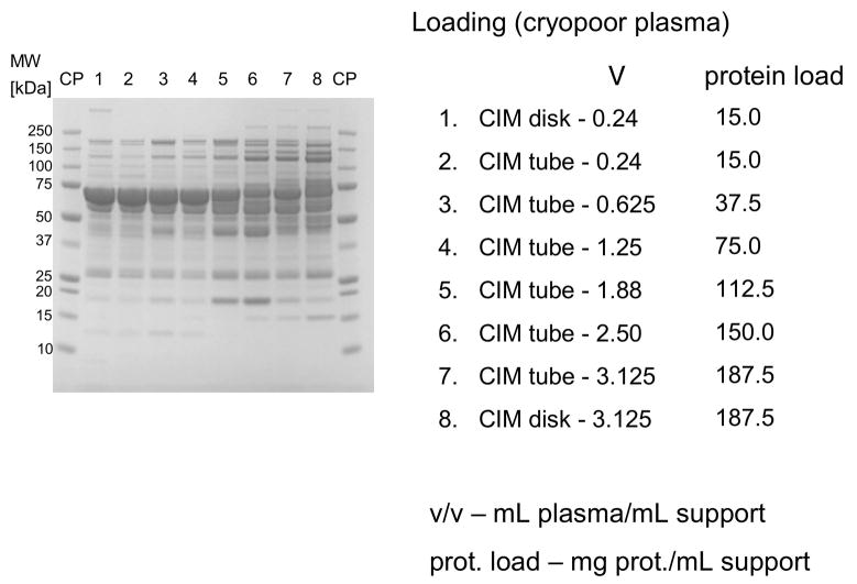
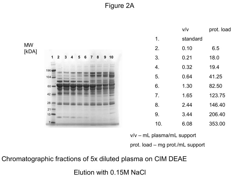
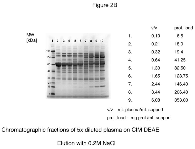
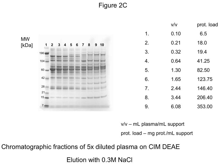
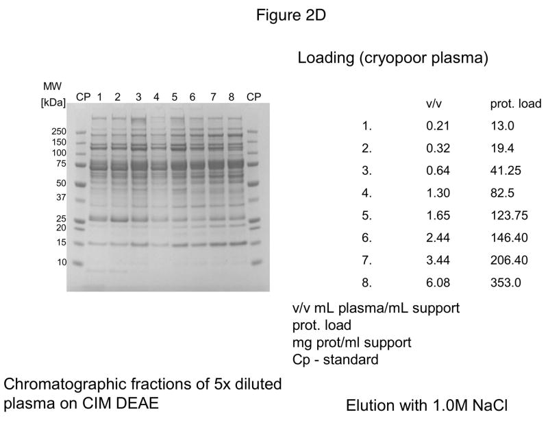
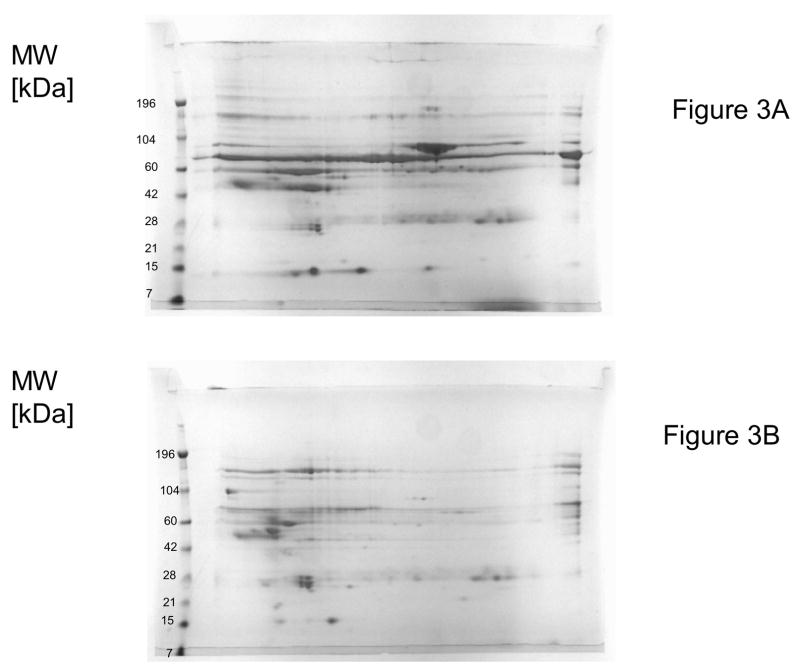
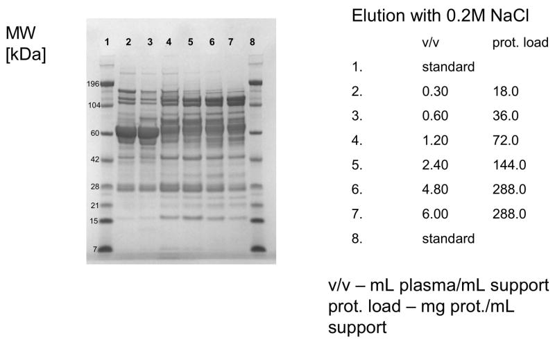
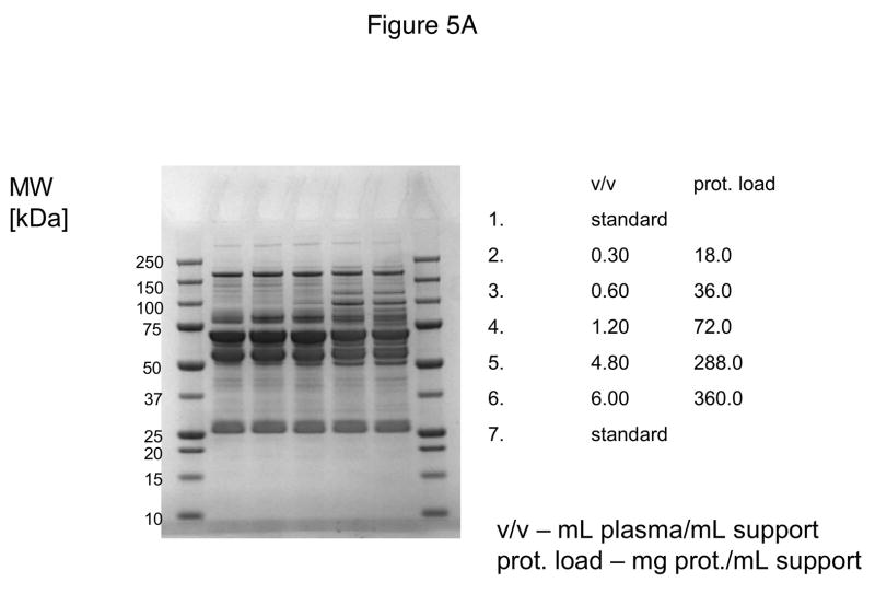
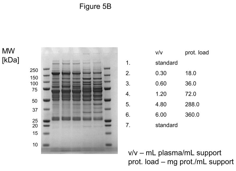
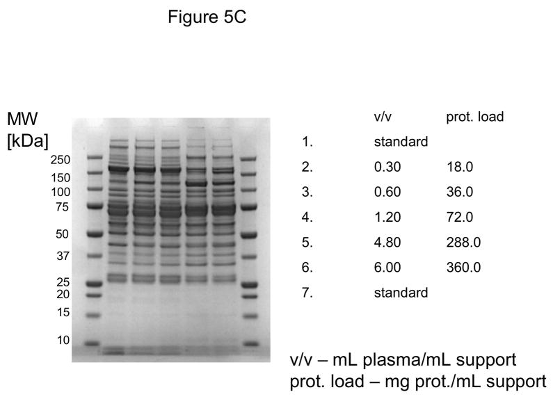
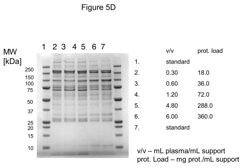
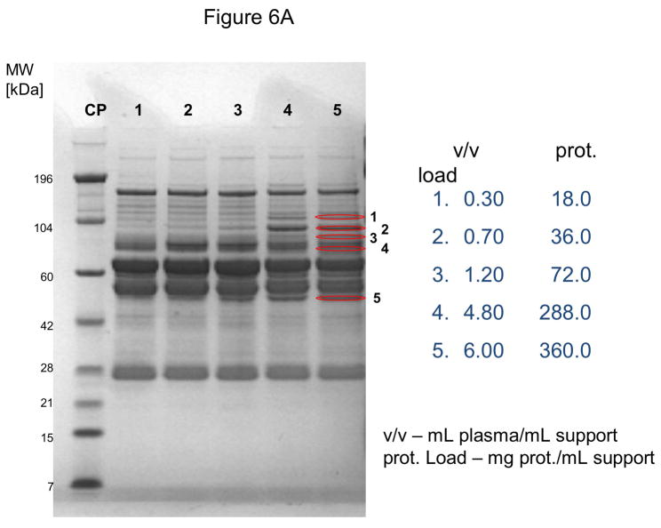
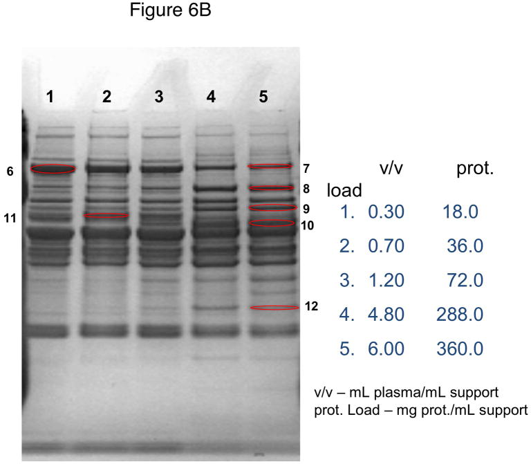
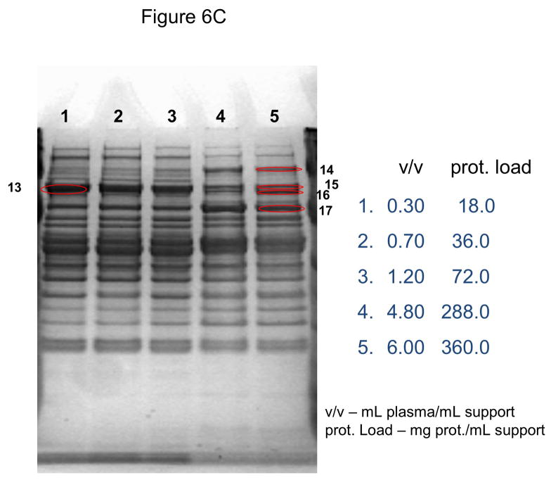
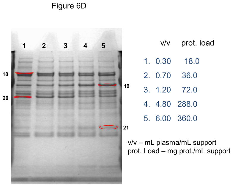
Human serum albumin (HSA) and immunoglobulin G (IgG) represent over 75% of all proteins present in human plasma. These two proteins frequently interfere with detection, determination and purification of low abundance proteins that can be potential biomarkers and biomarker candidates for various diseases. Some low abundance plasma proteins such as clotting factors and inhibitors are also important therapeutic agents. In this paper, the characterization of ion-exchange monolithic supports under overloading conditions was performed by use of sample displacement chromatography (SDC). If these supports were used for separation of human plasma, the composition of bound and eluted proteins in both anion- and cation-exchange mode is dependent on column loading. Under overloading conditions, the weakly bound proteins such as HSA in anion-exchange and IgG in cation-exchange mode are displaced by stronger binding proteins, and this phenomenon was not dependent on column size. Consequently, small monolithic columns with a column volume of 100 and 200 μL are ideal supports for high-throughput screening in order to develop new methods for separation of complex mixtures, and for sample preparation in proteomic technology.


Figures

Figure 1
Figure 1
Separation of proteins from human plasma on DEAE CIM monolithic supports under overloading conditions. CIM DEAE monolithic disk (column volume 340 μL) and tube (column volume 8 mL) were used. Human plasma was four times diluted with Buffer A, and bound proteins were eluted with 0.155 (A) by use of a step gradient. The flow rate was 2 mL/min for the CIM disk, and 10 mL/min for CIM tube. Collected fractions were analyzed by SDS-PAGE. For other separation conditions see Material and methods.
Figure 2
Figure 2
Separation of proteins from human plasma on DEAE weak anion-exchanger CIM monolithic supports under overloading conditions. CIM DEAE monolithic disk (column volume 340 μL) was used. Human plasma was four times diluted with Buffer A, and bound proteins were eluted with 0.155 (A), 0.2 (B), 0.3 (C) and 1 M NaCl (D)* by use of a step gradient. The flow rate was 2 mL/min. Collected fractions were analyzed by SDS-PAGE. For other separation conditions see Material and methods. *After injection of 37 μL plasma (6.5 mg protein/mL support), the amount of material eluted with 1M NaCl was not sufficient for subsequent SDS-PAGE
Figure 2
Figure 2
Separation of proteins from human plasma on DEAE weak anion-exchanger CIM monolithic supports under overloading conditions. CIM DEAE monolithic disk (column volume 340 μL) was used. Human plasma was four times diluted with Buffer A, and bound proteins were eluted with 0.155 (A), 0.2 (B), 0.3 (C) and 1 M NaCl (D)* by use of a step gradient. The flow rate was 2 mL/min. Collected fractions were analyzed by SDS-PAGE. For other separation conditions see Material and methods. *After injection of 37 μL plasma (6.5 mg protein/mL support), the amount of material eluted with 1M NaCl was not sufficient for subsequent SDS-PAGE
Figure 3
Figure 3
Two dimensional electrophoretic separation of fractions eluted with 0.155 M NaCl from CIM DEAE disk-shaped monolithic column (column volume 340 μL) and displacement of HSA under overloading conditions. A–0.25 mL plasma containing about 15 mg protein was loaded on the column (for comparison, see lane No. 1 in Figure 1A); B–1.06 mL plasma containing 187.5 mg protein was loaded on the column (for comparison, see lane No. 8 in Figure 1A). For separation conditions, see Figure 1 and Material and methods.
Figure 4
Figure 4
Separation of proteins from human plasma QA strong anion-exchanger CIM monolithic supports under overloading conditions. CIM QA monolithic disk (column volume 340 μL) was used. Human plasma was four times diluted with Buffer A, and bound proteins were eluted with 0.2, 0.3, 0.5 and 1 M NaCl by use of a step gradient. The flow rate was 2 mL/min. Collected fractions were analyzed by SDS-PAGE. Only the profiles of fractions eluted with 0.2 M NaCl are shown. For other separation conditions see Material and methods.
Figure 5
Figure 5
Separation of proteins from human plasma SO3 strong cation-exchanger CIM monolithic supports under overloading conditions. CIM SO3 monolithic disk (column volume 340 μL) was used. Human plasma was four times diluted with Buffer A, and bound proteins were eluted with 0.2 (A), 0.3(B), 0.5(C) and 1 M (D) NaCl by use of a step gradient. The flow rate was 2 mL/min. Collected fractions were analyzed by SDS-PAGE. For other separation conditions see Material and methods.
Figure 5
Figure 5
Separation of proteins from human plasma SO3 strong cation-exchanger CIM monolithic supports under overloading conditions. CIM SO3 monolithic disk (column volume 340 μL) was used. Human plasma was four times diluted with Buffer A, and bound proteins were eluted with 0.2 (A), 0.3(B), 0.5(C) and 1 M (D) NaCl by use of a step gradient. The flow rate was 2 mL/min. Collected fractions were analyzed by SDS-PAGE. For other separation conditions see Material and methods.
Figure 6
Figure 6
Use of 100 μL monolithic SO3 strong cation-exchanger CIM disk for separation of proteins from human plasma under overloading conditions. Human plasma was four times diluted with Buffer A, and bound proteins were eluted with 0.2 (A), 0.3(B), 0.5(C) and 1 M (D) NaCl by use of a step gradient. The flow rate was 1 mL/min. Collected fractions were analyzed by SDS-PAGE. For other separation conditions see Material and methods. Band labeled in the Figures were excised, digested by trypsin and used for identification by LC-MS/MS. Some identified proteins are listed in Table 1.
Figure 6
Figure 6
Use of 100 μL monolithic SO3 strong cation-exchanger CIM disk for separation of proteins from human plasma under overloading conditions. Human plasma was four times diluted with Buffer A, and bound proteins were eluted with 0.2 (A), 0.3(B), 0.5(C) and 1 M (D) NaCl by use of a step gradient. The flow rate was 1 mL/min. Collected fractions were analyzed by SDS-PAGE. For other separation conditions see Material and methods. Band labeled in the Figures were excised, digested by trypsin and used for identification by LC-MS/MS. Some identified proteins are listed in Table 1.

References

    1. Nice EC, Rothacker J, Weinstock J, Lim L, Catimel B. J Chromatogr A. 2007;1168:190. - PubMed
    1. Anderson NL, Anderson NG. Mol Cell Proteomics. 2002;1:845. - PubMed
    1. Issaq HJ. Electrophoresis. 2001;22:3629. - PubMed
    1. Ivanov YD, Govorun VM, Bykov VA, Archakov AI. Proteomics. 2006;6:1399. - PubMed
    1. Burnouf T. J Chromatogr B. 1995;664:3. - PubMed

Publication types

MeSH terms

LinkOut - more resources