. 2011 Jan;134(Pt 1):171-182.
doi: 10.1093/brain/awq294.

A founder mutation in Anoctamin 5 is a major cause of limb-girdle muscular dystrophy

Affiliations

A founder mutation in Anoctamin 5 is a major cause of limb-girdle muscular dystrophy

Debbie Hicks et al. Brain. 2011 Jan.

Abstract

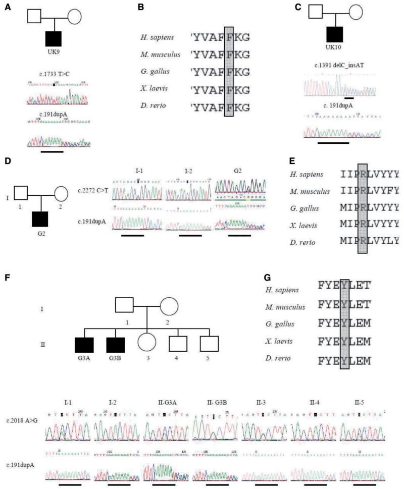
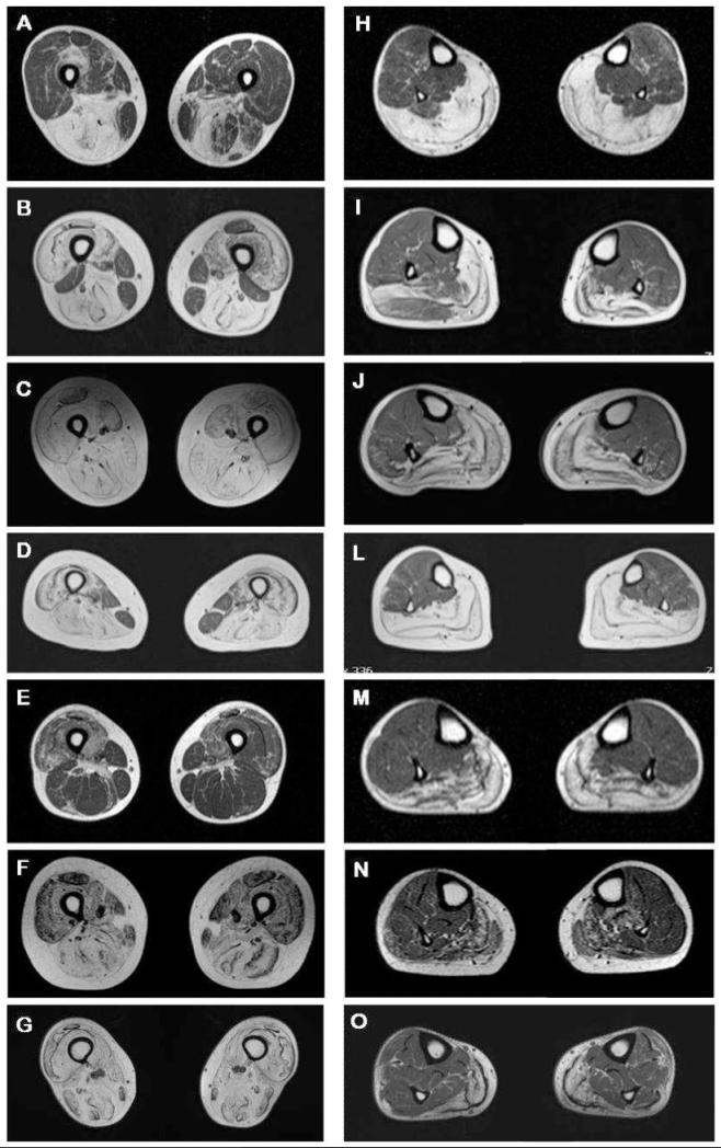
The limb-girdle muscular dystrophies are a group of disorders with wide genetic and clinical heterogeneity. Recently, mutations in the ANO5 gene, which encodes a putative calcium-activated chloride channel belonging to the Anoctamin family of proteins, were identified in five families with one of two previously identified disorders, limb-girdle muscular dystrophy 2L and non-dysferlin Miyoshi muscular dystrophy. We screened a candidate group of 64 patients from 59 British and German kindreds and found the truncating mutation, c.191dupA in exon 5 of ANO5 in 20 patients, homozygously in 15 and in compound heterozygosity with other ANO5 variants in the rest. An intragenic single nucleotide polymorphism and an extragenic microsatellite marker are in linkage disequilibrium with the mutation, suggesting a founder effect in the Northern European population. We have further defined the clinical phenotype of ANO5-associated muscular dystrophy. Patients show adult onset proximal lower limb weakness with highly raised serum creatine kinase values (average 4500 IU/l) and frequent muscle atrophy and asymmetry of muscle involvement. Onset varies from the early 20 s to 50 s and the weakness is generally slowly progressive, with most patients remaining ambulant for several decades. Distal presentation is much less common but a milder degree of distal lower limb weakness is often observed. Upper limb strength is only mildly affected and cardiac and respiratory function is normal. Females appear less frequently affected. In the North of England population we have identified eight patients with ANO5 mutations, suggesting a minimum prevalence of 0.27/100,000, twice as common as dysferlinopathy. We suggest that mutations in ANO5 represent a relatively common cause of adult onset muscular dystrophy with high serum creatine kinase and that mutation screening, particularly of the common mutation c.191dupA, should be an early step in the diagnostic algorithm of adult limb-girdle muscular dystrophy patients.


Figures

Figure 1
Figure 1
Identification of ANO5 mutations in UK and German families. Electrophoretograms of genomic DNA sequencing show heterozygous c.1733T > C p.Phe578Ser and c.191dupA mutations in patient UK9 (A), and protein alignment of residue Phe578 (B). Heterozygous c.1391delC_insAT and c.191dupA mutations in patient UK10 (C). Electrophoretograms of genomic DNA sequencing of patient G2 show heterozygous c.2272C > T (p.Arg758Cys) and c.191dupA mutations (D). His unaffected father carries the c.2272C > T mutation and unaffected mother carries the c.191dupA mutation. Protein alignment of residue Arg758 (E). Electrophoretograms of genomic DNA sequencing show heterozygous c.2018 A > G (p.Tyr673Cys) and c.191dupA mutations in patient G3A and G3B (F). Unaffected family members are carriers of either the c.2018A > G mutation (I-1, II-3, II-5) or for the c.191dupA mutation (I-2, II-4). Protein alignment of residue Tyr693 (G).
Figure 2
Figure 2
Analysis of two polymorphisms in or downstream of the ANO5 gene reveal a founder effect for the common mutation. Exons 1–22 are indicated by boxes, with non-coding regions unfilled. The intragenic SNP in exon 10 lies 29 Kb downstream of the site of the common mutation c.191dupA, and the extragenic microsatellite marker D11S1359 is located 135 Kb 3′ of ANO5 (A). A Fisher’s exact test of the frequency of rs7481951 and D11S1359 in mutant alleles (from patients homozygous for c.191dupA) and the allele frequency expected (for rs7481951); NCBI SNP database (http://www.ncbi.nlm.nih.gov/snp) or measured (for D11S1359) shows both polymorphisms to be in strong linkage disequilibrium with c.191dupA (B).
Figure 3
Figure 3
Clinical assessment of patients with ANO5 mutation. (A and B) Frontal and posterior view of the lower limbs of patient G3A showing atrophy of thighs and medial gastrocnemius and relative hypertrophy of lateral gastrocnemius. (C and D) Frontal and lateral view of the lower limbs of patient G2 showing severe atrophy of quadriceps and calves. (E) Focal atrophy of biceps muscles of patient UK12. (F, G and H) Severe hamstrings and quadriceps atrophy in patient UK3 and UK11. (I) Knee hyperextension in patient UK7A.
Figure 4
Figure 4
Atrophy of thigh and calf muscles assessed for seven patients by MRI. Section of thighs and calves for each patient are at the same level (A and H for patient UK1A; B and I for UK5A; C and J for UK5B; D and L for UK7A; E and M for UK11; F and N for G2; G and O for G3A) (A) Asymmetric atrophy of hamstrings in patient UK1A. (B) Severe atrophy of quadriceps and hamstrings muscle in patient UK5A with asymmetric sparing of rectus femoris, biceps femoris, gracilis and sartorius muscles. (C) Severe atrophy of thigh muscles in patient UK5B. (D) Severe atrophy of thigh muscles in patient UK7A with sparing of gracilis and sartorius muscles. (E) Moderate asymmetric patchy atrophy of quadriceps and hamstrings in patient UK11. (F and G) Moderate and severe generalized atrophy of the anterior and posterior thigh muscles in patient G2 and G3A. (H-O) Moderate to severe atrophy of the posterior compartment of the lower legs in all examined patients with atrophy of soleus and medial gastrocnemius and relative sparing of muscles of the anterior and lateral compartments, in particular tibialis anterior and posterior, peroneous longus and brevis as well as flexor digitorum longus and lateral gastrocnemius.
Figure 5
Figure 5
Histological, immunohistochemical and immunoblot findings in patients with ANO5 gene mutations. (A, B and C) H&E staining of muscle tissue from patients UK9, UK11 and UK5B showing myopathic or dystrophic changes. Notice the increase of internal nuclei (A), variation in fibre size, degeneration of muscle fibres (B) and increase in interfascicular and intrafascicular connective tissue (C). (D and E) neonatal myosin heavy chain labelling of muscle tissue from patient UK3 and UK11, respectively, indicating variable amounts of regenerating fibres. Brown fibres are undergoing regeneration. (F) WB analysis for dysferlin in patient UK9 showing a dysferlin band with normal size and amount. Equivalent protein loading was verified by Coomassie staining (blue band).

References

    1. Almaca J, Tian Y, Aldehni F, Ousingsawat J, Kongsuphol P, Rock JR, et al. TMEM16 proteins produce volume-regulated chloride currents that are reduced in mice lacking TMEM16A. J Biol Chem. 2009;284:28571–8. - PMC - PubMed
    1. Angelini C, Fanin M, Menegazzo E, Freda MP, Duggan DJ, Hoffman EP. Homozygous alpha-sarcoglycan mutation in two siblings: one asymptomatic and one steroid-responsive mild limb-girdle muscular dystrophy patient. Muscle Nerve. 1998;21:769–75. - PubMed
    1. Bolduc V, Marlow G, Boycott KM, Saleki K, Inoue H, Kroon J, et al. Recessive mutations in the putative calcium-activated chloride channel Anoctamin 5 cause proximal LGMD2L and distal MMD3 muscular dystrophies. Am J Hum Genet. 2010;86:213–21. - PMC - PubMed
    1. Fanin M, Pegoraro E, Matsuda-Asada C, Brown RH, Jr, Angelini C. Calpain-3 and dysferlin protein screening in patients with limb-girdle dystrophy and myopathy. Neurology. 2001;56:660–5. - PubMed
    1. Guglieri M, Straub V, Bushby K, Lochmüller H. Limb-girdle muscular dystrophies. Curr Opin Neurol. 2008;21:576–84. - PubMed

Publication types

MeSH terms