. 2011 Mar 4;286(9):7370-8.
doi: 10.1074/jbc.M110.187831. Epub 2010 Dec 25.

Ankyrin-B interactions with spectrin and dynactin-4 are required for dystrophin-based protection of skeletal muscle from exercise injury

Affiliations

Ankyrin-B interactions with spectrin and dynactin-4 are required for dystrophin-based protection of skeletal muscle from exercise injury

Gai Ayalon et al. J Biol Chem. .

Abstract

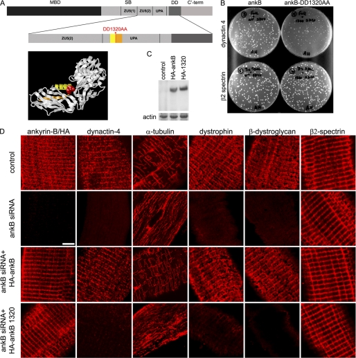
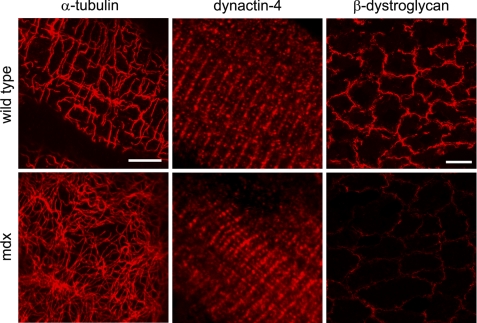
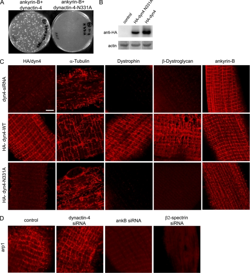
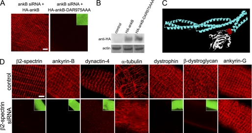
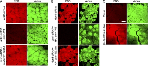
Costameres are cellular sites of mechanotransduction in heart and skeletal muscle where dystrophin and its membrane-spanning partner dystroglycan distribute intracellular contractile forces into the surrounding extracellular matrix. Resolution of a functional costamere interactome is still limited but likely to be critical for understanding forms of muscular dystrophy and cardiomyopathy. Dystrophin binds a set of membrane-associated proteins (the dystrophin-glycoprotein complex) as well as γ-actin and microtubules and also is required to align sarcolemmal microtubules with costameres. Ankyrin-B binds to dystrophin, dynactin-4, and microtubules and is required for sarcolemmal association of these proteins as well as dystroglycan. We report here that ankyrin-B interactions with β2 spectrin and dynactin-4 are required for localization of dystrophin, dystroglycan, and microtubules at costameres as well as protection of muscle from exercise-induced injury. Knockdown of dynactin-4 in adult mouse skeletal muscle phenocopied depletion of ankyrin-B and resulted in loss of sarcolemmal dystrophin, dystroglycan, and microtubules. Moreover, mutations of ankyrin-B and of dynactin-4 that selectively impaired binary interactions between these proteins resulted in loss of their costamere-localizing activity and increased muscle fiber fragility as a result of loss of costamere-associated dystrophin and dystroglycan. In addition, costamere-association of dynactin-4 did not require dystrophin but did depend on β2 spectrin and ankyrin-B, whereas costamere association of ankyrin-B required β2 spectrin. Together, these results are consistent with a functional hierarchy beginning with β2 spectrin recruitment of ankyrin-B to costameres. Ankyrin-B then interacts with dynactin-4 and dystrophin, whereas dynactin-4 collaborates with dystrophin in coordinating costamere-aligned microtubules.


Figures

FIGURE 1.
FIGURE 1.
Ankyrin-B requires dynactin-4 binding activity to promote association of microtubules, dystrophin, and dystroglycan with costameres. A, ankyrin-B domains (MBD, membrane binding domain; SB, spectrin binding domain; DD, death domain; ankB, ankyrin-B; C′-term, C′-terminal). The ankyrin-B-DD1320AA mutation (red) resides in an exposed loop in the UPA domain modeled from the UNC5b cytoplasmic domain structure (20). B, DD1320 is required for ankyrin-B-dynactin-4 but not β2 spectrin interaction in a yeast two-hybrid assay (as described under “Experimental Procedures”). C, representative immunoblot with HA antibody of homogenates of TA muscles depleted of endogenous ankyrin-B and transfected with either HA-ankyrin-B or HA-ankyrin-B-DD1320AA (as described under “Experimental Procedures”). D, single TA muscle fibers transfected in vivo with ankyrin-B siRNA and rescued with either wild type ankyrin-B or with ankyrin-B-DD1320AA (as described under “Experimental Procedures”). Rows (top to bottom), untransfected control, ankyrin-B siRNA (in both ankyrin-B staining is with ankyrin-B antibody), ankyrin-B siRNA co-transfected with HA-tagged ankyrin-B, and ankyrin-B siRNA co-transfected with HA-tagged ankyrin-B-DD1320AA (in both ankyrin-B staining is with an HA antibody). Scale bar, 5 microns.
FIGURE 2.
FIGURE 2.
Dystrophin is required for costamere patterning of microtubules but not for sarcolemmal association of microtubules and dynactin-4. Immunofluorescence labeling of muscle fibers (left and middle) and muscle cross-sections (right) of 5-week-old wild type, and mdx mice that lack dystrophin for localization of microtubules and dynactin-4. Left, grazing optical section at the level of the sarcolemma shows that in mdx mouse fibers, membrane-associated microtubules stained for α-tubulin are not organized in a costamere pattern, in contrast to in wild type fibers. Middle, dynactin-4 is localized along costameres in wild type as well as in mdx fibers. Right, cross-sections of TA muscles from wild type and mdx mice show loss of β-dystroglycan in the mdx muscle, establishing that neither dystrophin nor β-dystroglycan are required for costamere localization of dynactin-4 and for sarcolemmal association of microtubules. Scale bar for single fiber images (left and middle), 5 μm. Scale bar for muscle cross-sections (right), 20 μm.
FIGURE 3.
FIGURE 3.
Dynactin-4 requires ankyrin-B-binding activity for costamere localization of microtubules, dystrophin, and dystroglycan. A, N331A mutation of dynactin-4 impairs interaction with ankyrin-B (ankB) in yeast two-hybrid assays. B, shown are immunoblots (anti-HA) of HA-dynactin-4 and dynactin-4-N331A in transfected mouse TA muscles (as described under “Experimental Procedures”). C, single TA muscle fibers were transfected in vivo with a dynactin-4 siRNA and rescued with either wild type dynactin-4 or with dynactin-4-N331A (as described under “Experimental Procedures”). Rows from top to bottom, dynactin-4-siRNA-transfected muscle fibers (dynactin-4 detection with anti dynactin-4 antibody): middle row, dynactin-4 siRNA co-transfected with HA-wild type dynactin-4; bottom row, dynactin-4 siRNA cotransfected with HA-dynactin-4-N331A (middle and bottom rows, dynactin-4 detection with an anti-HA antibody). D, immunostaining for Arp1 in control, dynactin-4-, ankyrin-B-, and β2 spectrin-depleted muscle fibers (see “Experimental Procedures”). Scale bar, 5 microns.
FIGURE 4.
FIGURE 4.
Costamere association of ankyrin-B requires β2 spectrin. A, DAR975AAA mutation impairing spectrin binding prevents sarcolemmal localization of HA-ankyrin-B. Fibers were isolated and stained with an HA antibody. B, comparable expression levels (anti-HA immunoblot) of HA-ankyrin-B and HA-ankyrin-B-DAR975AAA in transfected TA muscles. D, β2 spectrin siRNA-transfected fibers exhibit loss of ankyrin-B (ankB), dystrophin, β-dystroglycan, dynactin-4, and costamere-associated microtubules while retaining ankyrin-G. Green insets show Venus expression in the knockdown fibers and cell boundaries. Scale bars, 5 microns.
FIGURE 5.
FIGURE 5.
Ankyrin-B-dynactin-4 interaction is required to maintain sarcolemmal integrity following exercise. Mice were transfected (Figs. 1, 3, and 4), exercised on a treadmill, and injected with EBD (see “Experimental Procedures”), and muscle sections evaluated for EBD uptake to assess sarcolemmal fragility. Red, EBD; Green, Venus, which marks the fibers that expressed the Venus vector containing siRNA. A, ankyrin-B (ankB)-depleted muscles exhibit EBD uptake and can be rescued by co-transfection with wild type HA-ankyrin-B but exhibit sarcolemmal fragility and EBD uptake when cotransfected with HA-ankyrin-B-DD1320AA, which is impaired in dynactin-4 binding. B, dynactin-4 knockdown results in sarcolemmal fragility and EBD uptake, which can be rescued by co-transfection with HA-dynactin-4 but are not with HA-dynactin-4-N331A, which is impaired in ankyrin-B binding. C, β2 spectrin knockdown results in sarcolemmal fragility and EBD uptake. Scale bar, 20 microns.

References

    1. Ervasti J. M. (2003) J. Biol. Chem. 278, 13591–13594 - PubMed
    1. Cohn R. D., Campbell K. P. (2000) Muscle Nerve 23, 1456–1471 - PubMed
    1. Dalkilic I., Kunkel L. M. (2003) Curr. Opin. Genet. Dev. 13, 231–238 - PubMed
    1. Craig S. W., Pardo J. V. (1983) Cell Motil. 3, 449–462 - PubMed
    1. Pardo J. V., Siliciano J. D., Craig S. W. (1983) Proc. Natl. Acad. Sci. U.S.A. 80, 1008–1012 - PMC - PubMed

Publication types

MeSH terms