. 2011 Jan;17(1):55-63.
doi: 10.1038/nm.2277. Epub 2010 Dec 26.

Receptor-mediated activation of ceramidase activity initiates the pleiotropic actions of adiponectin

Affiliations

Receptor-mediated activation of ceramidase activity initiates the pleiotropic actions of adiponectin

William L Holland et al. Nat Med. 2011 Jan.

Abstract

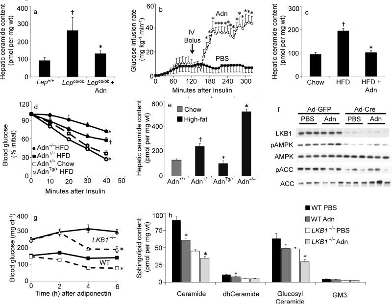
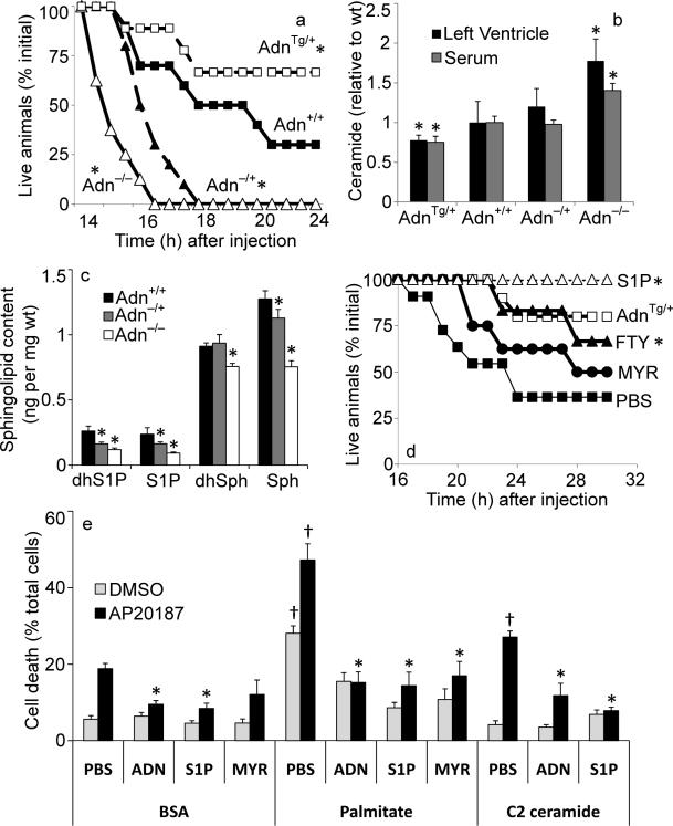
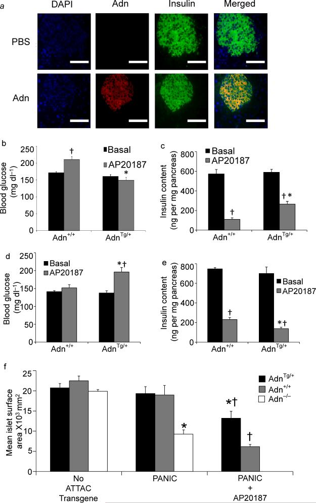
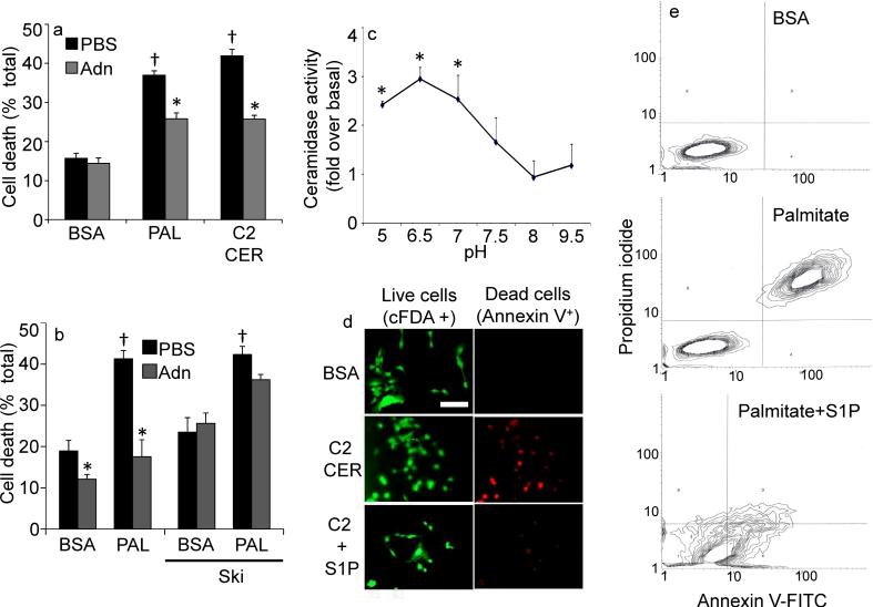
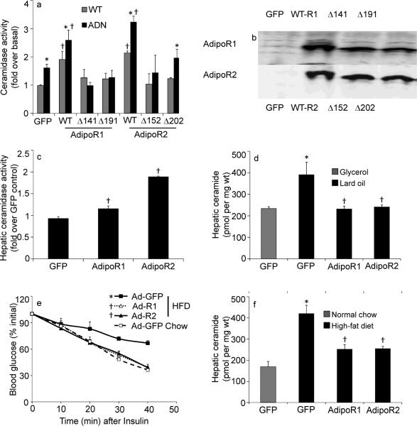
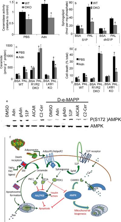
The adipocyte-derived secretory factor adiponectin promotes insulin sensitivity, decreases inflammation and promotes cell survival. No unifying mechanism has yet explained how adiponectin can exert such a variety of beneficial systemic effects. Here, we show that adiponectin potently stimulates a ceramidase activity associated with its two receptors, AdipoR1 and AdipoR2, and enhances ceramide catabolism and formation of its antiapoptotic metabolite--sphingosine-1-phosphate (S1P)--independently of AMP-dependent kinase (AMPK). Using models of inducible apoptosis in pancreatic beta cells and cardiomyocytes, we show that transgenic overproduction of adiponectin decreases caspase-8-mediated death, whereas genetic ablation of adiponectin enhances apoptosis in vivo through a sphingolipid-mediated pathway. Ceramidase activity is impaired in cells lacking both adiponectin receptor isoforms, leading to elevated ceramide levels and enhanced susceptibility to palmitate-induced cell death. Combined, our observations suggest a unifying mechanism of action for the beneficial systemic effects exerted by adiponectin, with sphingolipid metabolism as its core upstream signaling component.


Figures

Fig 1
Fig 1. Adiponectin rapidly lowers hepatic ceramide content and improves glucose homeostasis
(a) Total ceramide levels were quantified from liver of leptin deficient (ob/ob) mice after 60-minute treatments with full length adiponectin (Adn, 2 mg/kg, IV) or PBS (n=6/group). (b) Glucose infusion rates were calculated during hyperinsulinemic-euglycemic clamps peformed on conscious unrestrained ob/ob mice before and after a bolus of adiponectin (Adn, 2 mg/kg, IV) or PBS (n=5/group). (c) Total ceramide levels were quantified from liver of diet induced obese mice after 60-minute treatments with full length adiponectin (Adn, 2 mg/kg, IV) or PBS (n=9/group). (d–e) Adiponectin deficient (−/−), wildtype (+/+), or overexpressing mice (+/Tg) were maintained on high-fat diets (solid lines) or normal chow (dashed line) for 8 weeks prior to determination of (d) insulin tolerance and (e) hepatic ceramide content (n=7/group). (f–h) LKB1(fl/fl) mice were infected with adenovirus encoding either GFP or Cre recombinase 16 days prior to experiments (n=8/group). (f) Western blots of liver proteins probing against LKB1, phospho(T172)-AMPK, AMPK, phosphor(S79)-ACC1, and ACC1 (displayed in triplicate). (g) Whole blood glucose was monitored for 6 hours following injection of PBS (solid lines) or adiponectin (34 mg/kg, IV, dashed line). (h) Total hepatic sphingolipid levels were quantified by tandem MS/MS. * denotes significant effect of adiponectin (p<0.01). † Denotes significant effect of as compared to lean wildtype controls (p<0.05).
Fig 2
Fig 2. Adiponectin promotes cardiomyocyte and Heart ATTAC survival
(a) Female heart ATTAC transgenic mice crossed into indicated adiponectin backgrounds were challenged with AP20187 (0.010 μg/kg, IP) and survival was recorded as a Kaplan-Meier plot (n=12/group). (b) Ceramide was quantified from left ventricle or serum and normalized to the average content from adiponectin wildtype mice (63.9 pmol/mg in left ventricle, 9.5 pmol/μL in serum) (n=12/group). (c) Sphingosine, dihyrosphingosine, S1P, and dihydroS1P were quantified in left ventricle of WT (+/+), adiponectin (−/+), and adiponectin null mice (n=6/group). (d) Male HEART-ATTAC mice were treated with myriocin (0.3 mg/kg, IP), FTY720 (1 mg/kg, IP), S1P (1mg/kg, IP) or PBS immediately prior to injection with AP20187 (0.05 μg/kg, IP) and survival was recorded as a Kaplan-Meier plot (n=10/group). Additionally, treating HEART-ATTAC mice with S1P (1mg/kg, IP) just prior to AP20187 treatment prevented death in 100% of the animals tested (n=15) (e) Primary cardiomyocytes were isolated from heart ATTAC transgenic pups. After 72 hours of maintenance, cells were washed and treated with 2% BSA conjugated with: C2-ceramide (10 μM), myriocin (10 μM), palmitate (375 μM), or palmitate plus myriocin. PBS, adiponectin (3 μg/mL), or S1P (1 μM) were immediately added. Apoptosis was initiated by the addition of AP20187 (6.25 ng/mL), and viability was determined after 16 hours (n=6/group from 3 separate experiments). * denotes significant (p<0.05) difference from WT control. † denotes significant (p<0.01) effect of lipid treatment.
Fig 3
Fig 3. Adiponectin targets the endocrine pancreas and maintains β-cell mass
(a) Adiponectin, insulin, and nuclei were visualized by immunofluorescence after injection into adiponectin null mice (bar=100μm). (b) Random-fed blood glucose was assessed in male adiponectin transgenic vs. wildtype mice before and 10 days after treatment with AP20187 (0.2 mg/kg, IP, single injection) (n=12/group). (c) Total pancreatic insulin content was quantified from pancreas harvested 10 days after AP20187 treatment (n=6/group). (d–e) Female adiponectin null and wildtype PANIC-ATTAC mice were evaluated 10 days after initiating treatment with AP20187 (0.2 mg/kg, IP, twice daily for 3 days) or vehicle. (d) Random fed blood glucose was determined by glucometer (n=12/group). (e) Total pancreatic insulin content was quantified (n=6/group). (f) Pancreata were obtained 10 days after treatment with AP20187 (0.2 mg/kg) or vehicle from 10–12 week-old male mice overexpressing adiponectin (Tg/+), wildtype for adiponctin (+/+), or lacking adiponectin (−/−). Islet size was calculated by mean cross-sectional area of multicelled islets and reported as microns2/islet (n=6/condition). * denotes significant (p<0.02) difference between adiponectin transgenic (or adiponectin null) from WT animal of the same treatment. † denotes significant (p<0.02) effect of AP20187 treatment.
Fig 4
Fig 4. Adiponectin alters sensitivity to ceramide-induced apoptosis in INS-1 β-cells
(a) INS-1 cells were washed and removed to 2% BSA, Palmitate (750 μM in 2% BSA), or C2-Ceramide (50 μM in 2% BSA). Adiponectin (3 μg/mL) or PBS was immediately added and cell viability was determined after 18 hours (n=6/group from 3 separate experiments). (b) Cell viability was determined on INS-1 cells pretreated with sphingosine kinase inhibitor (2-(p-Hydroxyanilino)-4-(p-chlorophenyl) thiazole, 0.5 μM) or DMSO prior to delivery of adiponectin (3μg/mL) or PBS and maintained for 18 hours in the presence of 2% BSA or Palmitate (750 μM in 2% BSA) (c) Ceramidase activity was determined in lysates from cultured INS-1 cells under a range of pH conditions (n=4 from separate experiments) in the presence or absence of adiponectin (0.3 μg/mL, in vitro). “Fold change over baseline” refers to the change compared to BSA treatment without adiponectin. (d) INS-1 cells were challenged with C2-ceramide (50 μM) in the presence or absence of S1P (5 μM) cell death was determined by live/dead staining with cFDA or annexin V (image is representative of 3 separate experiments, bar=50μm). (e) Apoptosis of INS-1 cells was determined by FACS analysis of annexin V and propidium iodide stained cells following 18 hours of treatment with BSA, palmitate (750 μM), or coadministered palmitate and S1P (5μM) (representative of 3 independent experiments). * denotes significant (p<0.01) effect of adiponectin. † denotes significant (p<0.01) effect of pro-apoptotic insult.
Fig 5
Fig 5. Adiponectin Receptors 1 and 2 convey ceramidase activity in vivo
(a–b) HEK-293T cells were transiently cotransfected with GFP and murine AdipoR1, murine AdipoR2, GFP alone, or indicated point mutants for conserved histidine residues in AdipoR1 (H141R or H191R) or AdipoR2 (H152R or H202R). Ceramidase activity after in vitro treatment with adiponectin (0.3 μg/mL) (a) and protein expression (b) were assessed 48 hours after transfection (n=5 from separate experiments). (c–f) Hepatic overexpression of human AdipoR1, human AdipoR2, or GFP was accomplished by adenoviral delivery (0.5 ×10⁸ PFU/mouse). (c) Hepatic ceramidase activity was determined from fresh lysates 5 days after infection of 9 week-old wildtype mice C57/Bl6J mice (n=5/group). (d) Hepatic ceramides were measured following 6 hour infusion of 20% lard oil emulsions or fat-free glycerol control emulsions (n=6/group). (e–f) After 8 weeks of maintenance on high-fat diets, wildtype FVB mice were infected with AdipoR1, AdipoR2, or GFP. Insulin tolerance (e) and hepatic ceramide content (f) were determined 8 days after infection (n=6–8/group). *denotes significant (p<0.05) effect of adiponectin or lipid administration. † denotes significant (p<0.02) effect of genetic overexpression.
Fig 6
Fig 6. Ablating Adiponectin Receptors 1 and 2 impairs ceramidase activity, S1P generation and cell survival
(a) Ceramidase activity was determined from WT or AdipoR1/AdipoR2 double knockout (DKO) MEFs in the presence or absence of adiponectin (0.3 μg/mL, in vitro) (n=4). (b) S1P, and dihydroS1P were quantified from WT, or AdipoR1/AdipoR2 DKO MEFs after 12-hour incubation in palmitate (750 μM in 2% BSA) or BSA (2%) (n=6). (c) Ceramide levels were quantified from WT, AdipoR1/AdipoR2 DKO, or LKB1 knockout MEFs after 12-hour incubations with palmitate (750 μM in 2% BSA) or BSA (2%) supplemented with adiponectin (5 μg/mL) or PBS (n=6 from 3 separate experiments). (d) Cell viability was assessed in MEFs treated as in Fig. 6C after 16 hours of palmitate treatment. (n=5). (e) 60 minutes after removal from serum, cultured INS-1 cells were pretreated for 5 minutes with D-e-MAPP (100nM) or DMSO then treated for 10 minutes with full length adiponectin (Adn, 3μg/mL), truncated globular adiponectin (gAdn, 1 μg/mL), S1P (5 μM), AICAR (1 mM), or C2-ceramide (50 μM). Total and phosphorylated AMPK were probed by western blot (representative of 4 independent experiments). (f) Ceramide promotes apoptosis by aiding death receptor clustering, apoptosome formation, and Bax translocation. Ceramide impairs Akt activation via activation of PKCζ or PP2A. Adiponectin promotes the deacylation of ceramide by activating adiponectin receptors. The resulting sphingosine and S1P increase intracellular calcium and activate AMPK via stimulation of CAMKK. These actions promote survival, nutrient uptake, nutrient utilization and mitochondrial proliferation. * denotes significant (p<0.05) effect of adiponectin. † denotes p<0.05 compared to WT cells.

Comment in

References

    1. Yamauchi T, et al. Cloning of adiponectin receptors that mediate antidiabetic metabolic effects. Nature. 2003;423:762–769. - PubMed
    1. Combs TP, et al. A transgenic mouse with a deletion in the collagenous domain of adiponectin displays elevated circulating adiponectin and improved insulin sensitivity. Endocrinology. 2004;145:367–383. - PubMed
    1. Kim JY, et al. Obesity-associated improvements in metabolic profile through expansion of adipose tissue. J Clin Invest. 2007;117:2621–2637. - PMC - PubMed
    1. Holland WL, Summers SA. Sphingolipids, insulin resistance, and metabolic disease: new insights from in vivo manipulation of sphingolipid metabolism. Endocr Rev. 2008;29:381–402. - PMC - PubMed
    1. Takabe K, Paugh SW, Milstien S, Spiegel S. “Inside-out” signaling of sphingosine-1-phosphate: therapeutic targets. Pharmacol Rev. 2008;60:181–195. - PMC - PubMed

Publication types

MeSH terms

LinkOut - more resources