. 2011 Feb 20;333(2):151-9.
doi: 10.1016/j.mce.2010.12.027. Epub 2010 Dec 25.

Aryl hydrocarbon receptor activation in lactotropes and gonadotropes interferes with estradiol-dependent and -independent preprolactin, glycoprotein alpha and luteinizing hormone beta gene expression

Affiliations

Aryl hydrocarbon receptor activation in lactotropes and gonadotropes interferes with estradiol-dependent and -independent preprolactin, glycoprotein alpha and luteinizing hormone beta gene expression

Jinyan Cao et al. Mol Cell Endocrinol. .

Abstract

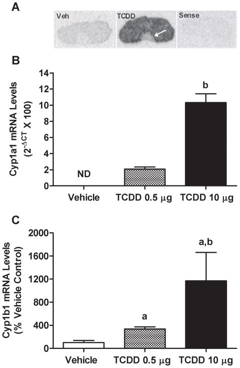
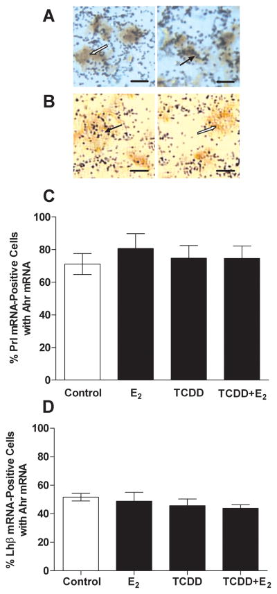
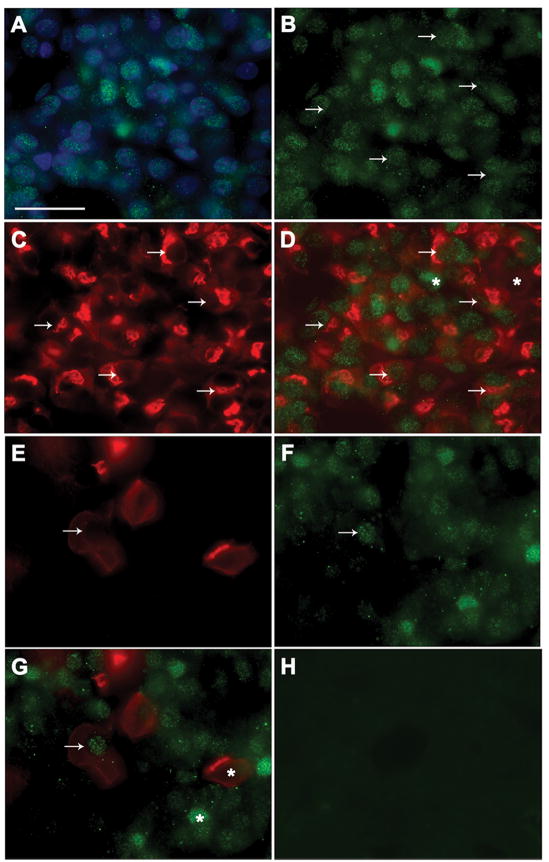
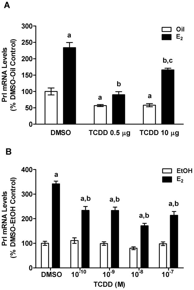
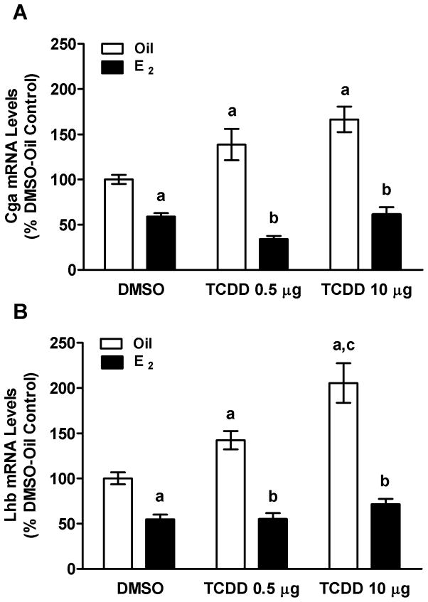
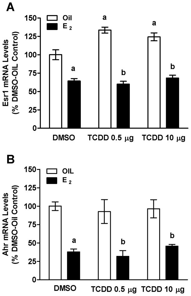
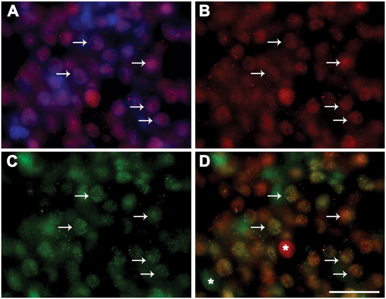
Arylhydrocarbon receptor (Ahr) activation by 2,3,7,8-tetrachlordibenzo-p-dioxin (TCDD) interferes with female reproductive functions, but there is little information on the specific targets of TCDD in the hypothalamic-pituitary-gonadal (HPG) axis. In these studies, we found that TCDD upregulated known AhR target genes, cytochrome p450 1a1 (Cyp1a1), Cyp1a2 and Cyp1b1 in the rat pituitary gland. Moreover, 75% of pituitary lactotropes and 45% of gonadotropes contained Ahr mRNA, and most Ahr-containing cells were estrogen receptor 1 (Esr1)-positive. TCDD abrogated estradiol (E(2))-induced prolactin (Prl) expression in vivo and in vitro; conversely, E(2) blocked TCDD upregulation of luteinizing hormone beta (Lhb) and glycoprotein hormone alpha polypeptide (Cga) expression. TCDD had no effect on levels of Ahr mRNA, but upregulated Esr1 mRNA. E(2) independently repressed Ahr and Esr1 expression and blocked TCDD upregulation of Esr1. Thus, complex interactions between Ahr and Esr alter Prl and luteinizing hormone (LH) synthesis by direct actions in lactotropes and gonadotropes. These findings provide important insights into how TCDD disrupts female reproductive functions.


Figures

FIG. 1
FIG. 1
Effects of TCDD on Cyp1a2, 1a1 and 1b1 gene expression in the pituitary gland in vivo. (A) Film autoradiogram of ISH using 35S-labeled cRNA probes for Cyp1a2 mRNA in pituitaries from OVX rats treated p.o. with TCDD 10 μg/kg bw or DMSO vehicle. White arrow indicates particularly intense signal in the intermediate lobe. QPCR measurements of Cyp1a1 (B) or Cyp1b1 (C) mRNA levels in OVX rats treated with either 0.5 or 10 μg/kg bw TCDD p.o. Bars represent mean ± SEM of 4–6 samples analyzed in duplicate. ND indicates no detectable signal in vehicle treatment group. aSignificantly different from vehicle-treated group; bsignificantly different from 0.5 μg group. Differences were considered significant if p<0.05.
FIG. 2
FIG. 2
Results of ISH studies colocalizing Ahr and Prl or Lhb mRNAs in pituitary glands of rats treated with E2, TCDD or both. Photomicrographs in the top panel show results of dual-label ISH with digoxigenin-labeled cRNA probes for (A) Prl mRNA (brown signal) or (B) Lhb mRNA (brown signal) and 35S-labeled cRNA probes for Ahr mRNA (black silver grains) hybridized to pituitary cryosections. White arrows denote cells containing either Prl mRNA (A) or Lhb mRNA (B) only. Black arrows indicate cells with both Ahr and Prl mRNAs (A) or Ahr and Lhb mRNAs (B). Scale bars = 5 μm. Graphs in the bottom panels show effects of TCDD and E2 on the percentages of lactotropes (C) and gonadotropes (D) that contained Ahr mRNA. Bars represent mean ± SEM.
Fig. 3
Fig. 3
Results of ICC studies colocalizing Ahr and Prl or Lhb proteins in rat anterior pituitary gland. (A-D) Photomicrographs of Ahr-ir in Hoeschst 33258-stained nuclei (A); cells with Ahr-ir (B, green), Prl-ir (C, red) or both Prl-ir and Ahr-ir (D). (E-H) Photomicrographs of cells with Lhb-ir (E, red), Ahr-ir (F, green), or both Lhb-ir with Ahr-ir (G); (H) example of negative control in which primary antibody was omitted. Arrows indicate dual-labeled cells and asterisks denote cells that express only Ahr-ir (D and G, green) or only Prl-ir (D, red around unstained nucleus) or Lhb-ir (G, red). Scale bar, 25 μm
FIG. 4
FIG. 4
Effects of E2 and TCDD on Prl mRNA levels in pituitaries of OVX rats in vivo and in GH3 cells in vitro. (A) QPCR measurements of Prl mRNA from rats OVX rats treated with Oil or E2 and DMSO or TCDD as described in Materials and Methods. Bars represent mean ± SEM of values from 5–6 animals run in duplicate. aSignificantly different from Oil-DMSO vehicle control; bsignificantly different from E2-DMSO group; csignificantly different from E2-TCDD 0.5 μg/kg bw group. (B) GH3 cells treated with 10−8 M E2 or ethanol (EtOH) and various concentrations of TCDD. Bars represent mean ± SEM of 10–11 samples each run in duplicate. aSignificantly different from corresponding EtOH control; bsignificantly different from E2-DMSO group. Differences were considered significant if p<0.05.
FIG. 5
FIG. 5
Effects of E2 and TCDD (0.5 or 10 μg/kg bw) or Oil or DMSO vehicle on Cga (A) and Lhb (B) mRNA levels in pituitary glands of OVX rats (See Materials and Methods for details of treatments). Bars represent mean ± SEM of values from 5–7 animals run in duplicate. aSignificantly different from Oil-DMSO vehicle control; bsignificantly different from corresponding Oil control; csignificantly different from Oil-TCDD 0.5 μg group. Differences were considered significant if p<0.05.
FIG. 6
FIG. 6
Effects of E2 and TCDD on Esr1 (A) or Ahr (B) mRNA levels in the pituitary glands of OVX rats measured using QPCR. Bars represent mean ± SEM of values from 5–7 animals run in duplicate. aSignificantly different from Oil-DMSO vehicle control; bsignificantly different from corresponding Oil control. Differences were considered significant if p<0.05.
FIG. 7
FIG. 7
Colocalization of Ahr and Esr1 proteins in rat anterior pituitary gland using ICC and Hoeschst 33258 to counterstain cell nuclei (blue, Panel A). Photomicrographs show Esr1-ir and Hoeschst 33258 stain (A), Esr1-ir (red; Panel B); Ahr-ir (green; Panel C) and colocalization of Esr1-ir with Ahr-ir (D). Arrows denote cells that contain both Esr1-ir and Ahr-ir; asterisks in (D) denote cells that express only Ahr-ir (green) or Esr1 (red). Scale bar, 25μm.

References

    1. Augustine RA, Kokay IC, Andrews ZB, Ladyman SR, Grattan DR. Quantitation of prolactin receptor mRNA in the maternal rat brain during pregnancy and lactation. J Mol Endocrinol. 2003;31:221–232. - PubMed
    1. Beischlag TV, Perdew GH. ER alpha-AHR-ARNT protein-protein interactions mediate estradiol-dependent transrepression of dioxin-inducible gene transcription. J Biol Chem. 2005;280:21607–21611. - PubMed
    1. Bestervelt LL, Pitt JA, Piper WN. Evidence for Ah receptor mediation of increased ACTH concentrations in primary cultures of rat anterior pituitary cells exposed to TCDD. Toxicol Sci. 1998;46:294–299. - PubMed
    1. Birnbaum LS. Endocrine effects of prenatal exposure to PCBs, dioxins, and other xenobiotics: implications for policy and future research. Environ Health Perspect. 1994;102:676–679. - PMC - PubMed
    1. Burbach KM, Poland A, Bradfield CA. Cloning of the Ah-receptor cDNA reveals a distinctive ligand-activated transcription factor. Proc Natl Acad Sci USA. 1992;89:8185–8189. - PMC - PubMed

Publication types

MeSH terms