. 2011 Mar 18;286(11):9150-61.
doi: 10.1074/jbc.M110.182428. Epub 2010 Dec 27.

Molecular control of polyene macrolide biosynthesis: direct binding of the regulator PimM to eight promoters of pimaricin genes and identification of binding boxes

Affiliations

Molecular control of polyene macrolide biosynthesis: direct binding of the regulator PimM to eight promoters of pimaricin genes and identification of binding boxes

Javier Santos-Aberturas et al. J Biol Chem. .

Abstract

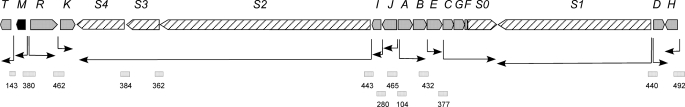
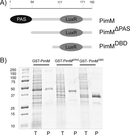
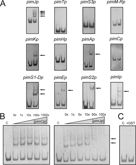
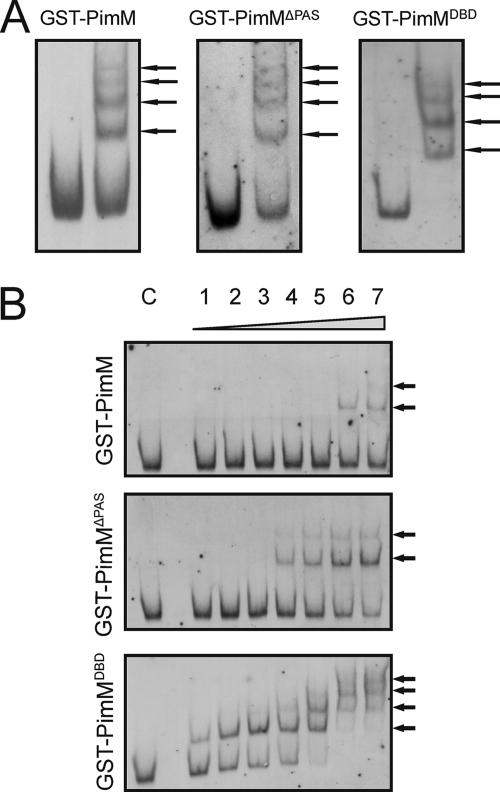
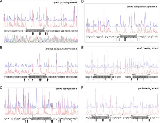
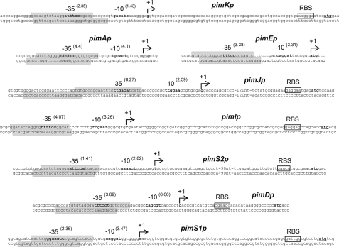
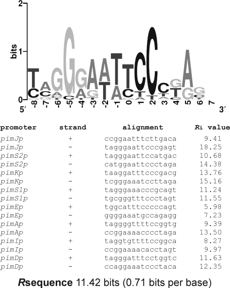
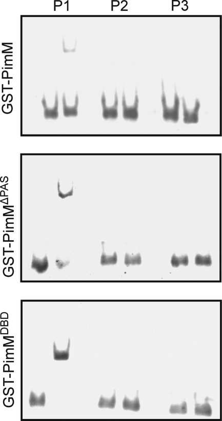
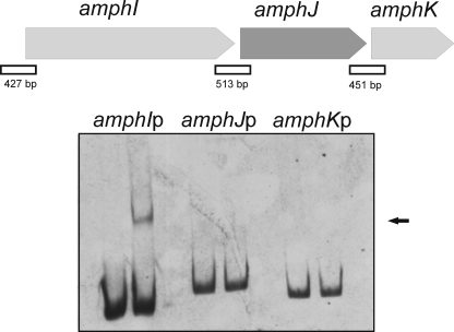
Control of polyene macrolide production in Streptomyces natalensis is mediated by the transcriptional activator PimM. This regulator, which combines an N-terminal PAS domain with a C-terminal helix-turn-helix motif, is highly conserved among polyene biosynthetic gene clusters. PimM, truncated forms of the protein without the PAS domain (PimM(ΔPAS)), and forms containing just the DNA-binding domain (DBD) (PimM(DBD)) were overexpressed in Escherichia coli as GST-fused proteins. GST-PimM binds directly to eight promoters of the pimaricin cluster, as demonstrated by electrophoretic mobility shift assays. Assays with truncated forms of the protein revealed that the PAS domain does not mediate specificity or the distinct recognition of target genes, which rely on the DBD domain, but significantly reduces binding affinity up to 500-fold. Transcription start points were identified by 5'-rapid amplification of cDNA ends, and the binding regions of PimM(DBD) were investigated by DNase I protection studies. In all cases, binding took place covering the -35 hexamer box of each promoter, suggesting an interaction of PimM and RNA polymerase to cause transcription activation. Information content analysis of the 16 sequences protected in target promoters was used to deduce the structure of the PimM-binding site. This site displays dyad symmetry, spans 14 nucleotides, and adjusts to the consensus TVGGGAWWTCCCBA. Experimental validation of this binding site was performed by using synthetic DNA duplexes. Binding of PimM to the promoter region of one of the polyketide synthase genes from the Streptomyces nodosus amphotericin cluster containing the consensus binding site was also observed, thus proving the applicability of the findings reported here to other antifungal polyketides.


Figures

FIGURE 1.
FIGURE 1.
Organization of the pimaricin gene cluster. The pointed boxes indicate the direction of transcription. The pimM gene is indicated in black, and the polyketide synthase genes are shown as striped pointed boxes. The arrows indicate deduced transcriptional units. Square boxes, DNA fragments used in mobility shift experiments (sizes (bp) are indicated below).
FIGURE 2.
FIGURE 2.
Purification of GST fusion proteins in E. coli BL21. A, scheme of PimM and its truncated versions. Numbers indicate amino acid residues from the N terminus. B, purification of GST-PimM, GST-PimMΔPAS, and GST-PimMDBD proteins by affinity chromatography on glutathione-Sepharose. Lanes T, total E. coli cell extract; lanes P, purified proteins after affinity chromatography. Left lane, molecular size markers (in kDa).
FIGURE 3.
FIGURE 3.
GST-PimM DNA binding assay results. A, EMSA of GST-PimM binding to different putative promoter regions. Left lane, control without protein; right lane, 60 μm GST-PimM protein. The pimJ promoter region was used as a positive control. The arrows indicate the DNA-protein complexes. Promoter names are indicated above the pictures. All experiments were carried out with 2 ng of labeled DNA probe. B, left panel, competition experiment between pimJp and pimCp. Note that 1000-fold higher concentrations of unlabeled pimCp competitor DNA failed to decrease the intensities of the pimJp retardation bands. Right panel, competition experiment between labeled pimJp and unlabeled pimJp. Both experiments were performed with 19 μm GST-PimM protein. C, control reaction with the pimJ promoter region and 60 μm pure GST protein. In all cases, lane C indicates control without protein.
FIGURE 4.
FIGURE 4.
GST-PimMDBD binds promoters with highest affinity. Analysis by EMSA of the binding of the different GST fusion proteins to the pimJ promoter. A, binding of the different GST fusion proteins in a protein-saturated assay (60 μm). B, lane C, control without protein; lane 1, 19 nm protein; lane 2, 95 nm protein; lane 3, 190 nm protein; lane 4, 950 nm protein; lane 5, 1.9 μm protein; lane 6, 9.5 μm protein; lane 7, 19 μm protein. The arrows indicate the DNA-protein complexes. Top, GST-PimM; middle, GST-PimMΔPAS; bottom, GST-PimMDBD.
FIGURE 5.
FIGURE 5.
Identification of binding sites. DNase I footprints of the GST-PimMDBD protein bound to the promoter regions of pimS2 (A and B), pimJ (C and D), and pimS1-D (E and F). In each panel, the upper electropherogram (blue line) shows the control reaction. The protected nucleotide sequence is boxed; hypersensitive sites (arrows) are also indicated. Sequencing reactions are not included except in A. Coordinates are from the transcriptional start point. In the case of the bidirectional promoter pimS1-Dp, the gene to which coordinates refer is indicated in parentheses.
FIGURE 6.
FIGURE 6.
Transcriptional start site of promoters regulated by PimM and protected regions. The position of the transcriptional start site was determined by 5′-RACE. The putative −10 and −35 hexanucleotides are in boldface type. Scores resulting from the comparison with the matrices reported by Bourn and Babb (28) for Streptomyces are indicated in parentheses. The TSP is indicated by a bent arrow and boldface type. Nucleotides showing homology with the 16 S RNA, which could form a ribosome-binding site, are framed with a box labeled RBS. The start codon is shown in boldface type and underlined. Protected nucleotide sequences are indicated with shaded boxes. Note that pimA and pimE are transcribed as leaderless mRNAs, thus lacking RBS.
FIGURE 7.
FIGURE 7.
Sequence logo of PimM binding site. Sequence logo of the nucleotides sequences that constitute the PimM binding site. The logo was constructed with the 16 sequences shown to be protected by PimM binding in footprinting experiments. The height of each letter is proportional to the frequency of the base, and the height of the letter stack shows the conservation in bits at that position (33). Note the high conservation of the eight central positions. The total information (Rsequence) for the binding site is 11.42 bits (0.71 bits/base). An alignment of the 16 binding sites is indicated below. Information contents (Ri) of each binding site are shown.
FIGURE 8.
FIGURE 8.
Validation of PimM binding site. Shown is analysis by EMSA of the binding of the different GST fusion proteins to synthetic duplexes. Base composition of the duplexes is indicated under “Experimental Procedures.” Left lane, control without protein; right lane, 9 μm protein. Top, GST-PimM; middle, GST-PimMΔPAS; bottom, GST-PimMDBD. Note that only the P1 duplex, with the consensus binding site, forms a complex with the proteins.
FIGURE 9.
FIGURE 9.
GST-PimM binds the amphI promoter. Shown is analysis by EMSA of GST-PimM binding to different putative promoter regions of some amphotericin genes. Left lane, control without protein; right lane, 60 μm GST-PimM protein. The arrow indicates the DNA-protein complex. Putative promoter names are indicated above the pictures. All experiments were carried out with 2 ng of labeled DNA probe. The organization of the studied genes in the S. nodosus chromosome is indicated above. Square boxes indicate DNA fragments used in mobility shift experiments.

References

    1. Bibb M. (1996) Microbiology 142, 1335–1344 - PubMed
    1. Aparicio J. F., Mendes M. V., Antón N., Recio E., Martín J. F. (2004) Curr. Med. Chem. 11, 1645–1656 - PubMed
    1. te Welscher Y. M., ten Napel H. H., Balagué M. M., Souza C. M., Riezman H., de Kruijff B., Breukink E. (2008) J. Biol. Chem. 283, 6393–6401 - PubMed
    1. Caffrey P., Aparicio J. F., Malpartida F., Zotchev S. B. (2008) Curr. Top. Med. Chem. 8, 639–653 - PubMed
    1. Mendes M. V., Recio E., Fouces R., Luiten R., Martín J. F., Aparicio J. F. (2001) Chem. Biol. 8, 635–644 - PubMed

Publication types

MeSH terms