Review
. 2011 Jan 15;124(Pt 2):161-70.
doi: 10.1242/jcs.064576.

The regulation of autophagy - unanswered questions

Affiliations
Review

The regulation of autophagy - unanswered questions

Yongqiang Chen et al. J Cell Sci. .

Abstract

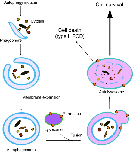
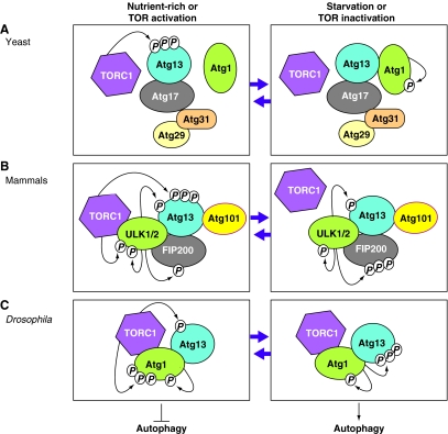
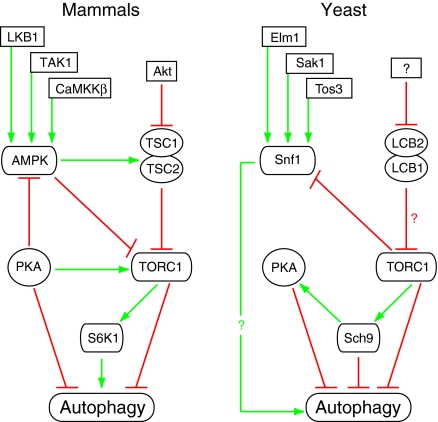
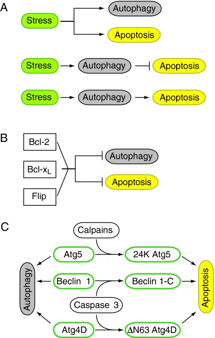
Autophagy is an intracellular lysosomal (vacuolar) degradation process that is characterized by the formation of double-membrane vesicles, known as autophagosomes, which sequester cytoplasm. As autophagy is involved in cell growth, survival, development and death, the levels of autophagy must be properly regulated, as indicated by the fact that dysregulated autophagy has been linked to many human pathophysiologies, such as cancer, myopathies, neurodegeneration, heart and liver diseases, and gastrointestinal disorders. Substantial progress has recently been made in understanding the molecular mechanisms of the autophagy machinery, and in the regulation of autophagy. However, many unanswered questions remain, such as how the Atg1 complex is activated and the function of PtdIns3K is regulated, how the ubiquitin-like conjugation systems participate in autophagy and the mechanisms of phagophore expansion and autophagosome formation, how the network of TOR signaling pathways regulating autophagy are controlled, and what the underlying mechanisms are for the pro-cell survival and the pro-cell death effects of autophagy. As several recent reviews have comprehensively summarized the recent progress in the regulation of autophagy, we focus in this Commentary on the main unresolved questions in this field.


Figures

Fig. 1.
Fig. 1.
The autophagy pathway and its roles in cell survival and cell death. In the presence of an autophagy inducer, cytoplasmic materials, such as protein aggregates and organelles, are sequestered by a pre-autophagosomal membrane structure, the phagophore. The phagophore membrane then expands and encloses its cargo to form a double-membrane vesicle, the autophagosome. The autophagosome fuses with a lysosome (or a vacuole in yeast) to form an autolysosome, in which the enclosed cargo is degraded by acid hydrolases. After the resulting macromolecules are transported back into the cytosol through membrane permeases, they can either be used to synthesize proteins or can be oxidized by the mitochondria to generate ATP for cell survival. However, when autophagy occurs at excessive levels or under certain physiological conditions it can lead to type II programmed cell death (type II PCD). See the text for additional details.
Fig. 2.
Fig. 2.
Dynamics of Atg1 complexes upon autophagy induction in different eukaryotes. (A) In yeast, under nutrient-rich conditions, the active TOR complex 1 (TORC1) hyperphosphorylates Atg13 (Kamada et al., 2010). This prevents the association of Atg1 with Atg13, which is bound to Atg17, Atg31 and Atg29, leading to inhibition of autophagy induction. Under starvation conditions when TORC1 is inactivated, Atg13 is no longer phosphorylated by TORC1, whereas Atg1 is autophosphorylated, leading to the association of Atg1 with the complex between Atg13, Atg17, Atg31 and Atg29, and subsequent autophagy induction (Cebollero and Reggiori, 2009; Chang and Neufeld, 2010; Kamada et al., 2010; Nakatogawa et al., 2009). (B) In contrast to yeast, mammalian ULK (ULK1 or ULK2, the homologs of yeast Atg1) forms a stable complex with mammalian Atg13, FIP200 (a putative counterpart of yeast Atg17) and Atg101 (an Atg13-binding protein), irrespective of TORC1 activation. Under nutrient-rich conditions, the active TORC1 associates with the ULK complex (ULK1 (or ULK2)–Atg13–FIP200-Atg101), phosphorylates ULK1 (or ULK2) and hyperphosphorylates Atg13, which inhibits the kinase activity of ULK1 (or ULK2) and thus blocks autophagy induction. Under starvation conditions when TORC1 is inactivated, TORC1 dissociates from the ULK complex, preventing phosphorylation of Atg13 and ULK1 (or ULK2) by TORC1 and leading to autophagy induction, whereas ULK1 (or ULK2) still phosphorylates Atg13 and itself, and hyperphosphorylates FIP200 (Chang and Neufeld, 2010; Mizushima, 2010; Yang and Klionsky, 2010). (C) Similar to the situation in mammals, in Drosophila Atg1 forms a complex with Atg13 irrespective of TORC1 activation (Chang and Neufeld, 2010). Under nutrient-rich conditions, the active TORC1 phosphorylates Atg13 and hyperphosphorylates Atg1, leading to the inhibition of autophagy induction. Under starvation conditions, when TORC1 is inactivated, Atg1 and Atg13 are no longer phosphorylated by TORC1, whereas Atg1 still phosphorylates itself and hyperphosphorylates Atg13, leading to autophagy induction. Figure modified from Chang and Neufeld (Chang and Neufeld, 2010) with permission.
Fig. 3.
Fig. 3.
The Atg8 and Atg12 ubiquitin-like conjugation systems in yeast. Atg8 (or the mammalian homolog LC3) is an ubiquitin-like (Ubl) protein that is cleaved by the cysteine protease Atg4 to expose a glycine residue that is then covalently conjugated with phosphatidylethanolamine (PE) (Ichimura et al., 2000). Atg12 is another Ubl protein that is covalently conjugated to Atg5 and the resulting Atg12–Atg5 conjugate is then attached to Atg16 to form a dimer of the trimeric Atg12–Atg5–Atg16 complex (Geng and Klionsky, 2008). Conjugation reactions of Atg12 and Atg8 are catalyzed by the E1-like enzyme Atg7 and the E2-like enzymes Atg10 (for conjugation of Atg12) and Atg3 (for Atg8) and result in the formation of either a multimeric complex with Atg5 (involving Atg12) or a lipid conjugate (with Atg8). In contrast to Atg5 conjugation to Atg12, Atg8 is conjugated to PE and not to a protein, thereby allowing membrane association. Furthermore, Atg4 cleaves the Atg8 (or LC3) precursor before conjugation and Atg8–PE (or LC3-II) during the subsequent deconjugation. Figure modified from Geng and Klionsky (Geng and Klionsky, 2008) with permission.
Fig. 4.
Fig. 4.
The TOR signaling pathways regulates autophagy in mammalian cells and yeast. In mammals, AMPK can be activated by its upstream factors LKB1, TAK1, and CaMKKβ (Yang and Klionsky, 2010). In yeast, Snf1 can be activated by its upstream factors Elm1, Sak1, and Tos3 (Hedbacker and Carlson, 2008). In yeast, the mammalian TSC1 and TSC2 homologs LCB2 and LCB1 (Gable et al., 2000) might form a complex that potentially inhibits TORC1, although this has not been experimentally demonstrated. A putative Akt homolog upstream of LCB2 and LCB1 also needs to be identified in yeast. Green arrows indicate interactions that induce autophagy, red bars indicate inhibition. See the text for details.
Fig. 5.
Fig. 5.
Crosstalk between autophagy and apoptosis in mammalian cells. (A) In a cell, the same stress induces both autophagy and apoptosis as independent processes (top). A stress can also induce autophagy, which then inhibits apoptosis (middle) or, alternatively, a stress induces autophagy, which can be the trigger of apoptosis (bottom). (B) Both autophagy and apoptosis are negatively regulated by Bcl-2, Bcl-xL and Flip. (C) Autophagy proteins Atg5, beclin 1 and Atg4D function in autophagy in their unmodified form, but also have a role in apoptosis after cleavage by either calpains, which target Atg5 and give rise to 24 K Atg5, or caspase 3, which cleaves beclin 1, resulting in a C-terminal fragment of beclin 1 (Beclin 1-C), and Atg4D, truncating it at the canonical caspase cleavage sequence (DEVD63K) to ΔN63 Atg4D.

References

    1. Alexander A., Cai S. L., Kim J., Nanez A., Sahin M., MacLean K. H., Inoki K., Guan K. L., Shen J., Person M. D., et al. (2010). ATM signals to TSC2 in the cytoplasm to regulate mTORC1 in response to ROS. Proc. Natl. Acad. Sci. USA 107, 4153-4158 - PMC - PubMed
    1. Armour S. M., Baur J. A., Hsieh S. N., Land-Bracha A., Thomas S. M., Sinclair D. A. (2009). Inhibition of mammalian S6 kinase by resveratrol suppresses autophagy. Aging 1, 515-528 - PMC - PubMed
    1. Baehrecke E. H. (2005). Autophagy: dual roles in life and death? Nat. Rev. Mol. Cell Biol. 6, 505-510 - PubMed
    1. Balgi A. D., Fonseca B. D., Donohue E., Tsang T. C., Lajoie P., Proud C. G., Nabi I. R., Roberge M. (2009). Screen for chemical modulators of autophagy reveals novel therapeutic inhibitors of mTORC1 signaling. PLoS ONE 4, e7124 - PMC - PubMed
    1. Bassham D. C. (2009). Function and regulation of macroautophagy in plants. Biochim. Biophys. Acta 1793, 1397-1403 - PubMed

Publication types

Substances