Comparative Study
. 2011 Apr;79(4):639-48.
doi: 10.1124/mol.110.069104. Epub 2010 Dec 27.

The high-affinity cAMP-specific phosphodiesterase 8B controls steroidogenesis in the mouse adrenal gland

Affiliations
Comparative Study

The high-affinity cAMP-specific phosphodiesterase 8B controls steroidogenesis in the mouse adrenal gland

Li-Chun Lisa Tsai et al. Mol Pharmacol. 2011 Apr.

Abstract

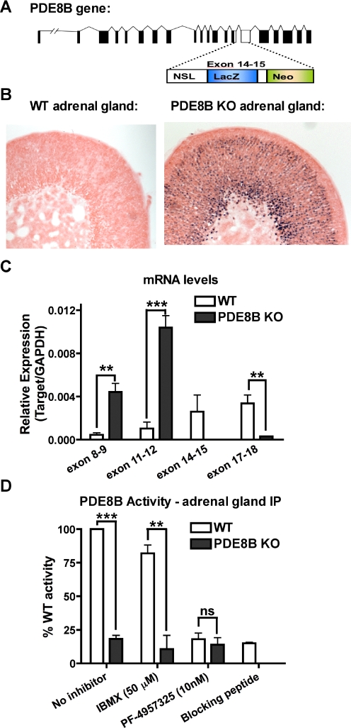
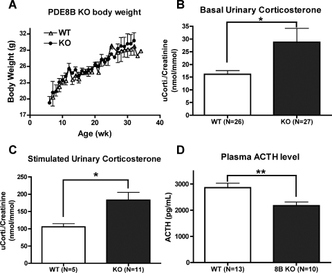
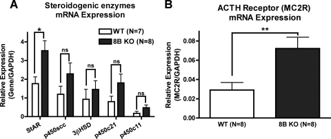
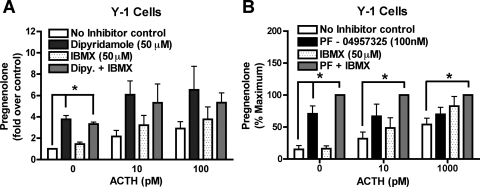
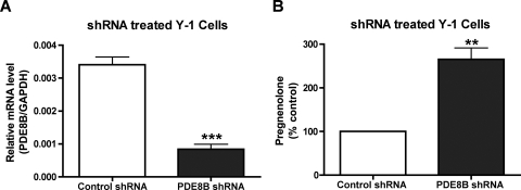
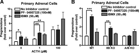
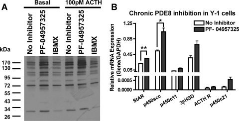
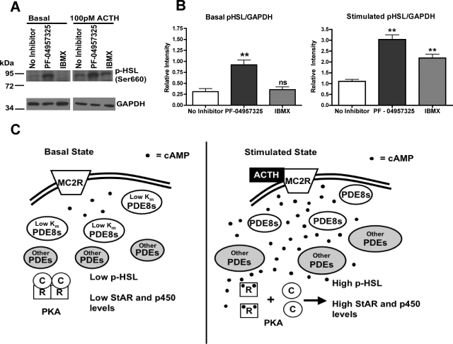
The functions of the phosphodiesterase 8B (PDE8) family of phosphodiesterases have been largely unexplored because of the unavailability of selective pharmacological inhibitors. Here, we report a novel function of PDE8B as a major regulator of adrenal steroidogenesis using a genetically ablated PDE8B mouse model as well as cell lines treated with either a new PDE8-selective inhibitor or a short hairpin RNA (shRNA) construct against PDE8B. We demonstrate that PDE8B is highly enriched in mouse adrenal fasciculata cells, and show that PDE8B knockout mice have elevated urinary corticosterone as a result of adrenal hypersensitivity toward adrenocorticotropin. Likewise, ablation of PDE8B mRNA transcripts by an shRNA construct potentiates steroidogenesis in the commonly used Y-1 adrenal cell line. We also observed that the PDE8-selective inhibitor (PF-04957325) potentiates adrenocorticotropin stimulation of steroidogenesis by increasing cAMP-dependent protein kinase activity in both primary isolated adrenocortical cells and Y-1 cells. It is noteworthy that PDE8s have their greatest control under low adrenocorticotropin-stimulated conditions, whereas other higher K(m) PDE(s) modulate steroidogenesis more effectively when cells are fully stimulated. Finally, both genetic ablation of PDE8B and long-term pharmacological inhibition of PDE8s cause increased expression of steroidogenic enzymes. We conclude that PDE8B is a major regulator of one or more pools of cAMP that promote steroidogenesis via both short- and long-term mechanisms. These findings further suggest PDE8B as a potential therapeutic target for the treatment of several different adrenal diseases.


Figures

Fig. 1.
Fig. 1.
PDE8B is highly expressed in AZF cells. A, the catalytic domain (exon 14–15) of PDE8B gene was disrupted by a construct containing a lacZ reporter gene, a neomycin resistance gene, and premature stop codons. B, X-gal staining of PDE8B KO adrenal gland showed that the promoter of PDE8B gene was active, and PDE8B was expressed in the AZF cells. C, the full-length PDE8B mRNA was not transcribed in the PDE8B KO adrenal. However, the 5′ mRNA region of PDE8B was up-regulated perhaps due to the absence of functional PDE8B (n = 3). D, the absence of functional PDE8B enzyme in PDE8B KO adrenals was shown by immunoprecipitating PDE8B from the PDE8B KO in comparison to the WT control (n = 3). The data are reported as means ± S.E.M., and the data were analyzed by Student's t test (unpaired, two-tailed): ns, no significance; **, p < 0.01; ***, p < 0.001.
Fig. 2.
Fig. 2.
PDE8B KO mice exhibit adrenal hypersensitivity in vivo. PDE8B KO mice had no gross developmental defects compared with their WT littermates. A, the PDE8B KO mice maintained normal body weight under standard lab chow diet (n = 3–7). B, PDE8B KO mice had elevated basal urinary corticosterone (n = 26–27). C, PDE8B KO mice also exhibited increased stimulated corticosterone levels, when mice were mildly stressed via an intraperitoneal saline injection (n = 5–11). D, the circulating adrenocorticotropin (ACTH) level of the PDE8B KO was not higher than WT control (n = 10–13). The data are reported as means ± S.E.M., and the data were analyzed by Student's t test (unpaired, two-tailed): *, p < 0.05; **, p < 0.01.
Fig. 3.
Fig. 3.
PDE8B gene ablation increases mRNA expressions of StAR protein and MC2R. The long-term phase of steroidogenesis elicits cAMP-dependent transcriptional activation to increase steroid production. A, PDE8B KO adrenals had an increase of mRNA level of steroidogenic enzyme, StAR protein (n = 7–8). B, the mRNA of the adrenocorticotropin (ACTH) receptor (MC2R), which is a known cAMP-activated transcript, was also elevated in PDE8B KO adrenals (n = 8). The data are reported as means ± S.E.M., and the data were analyzed by Student's t test (unpaired, two-tailed): ns, no significance; *, p > 0.05; **, p < 0.01.
Fig. 4.
Fig. 4.
PDE8 inhibition with inhibitors increases short-term adrenal steroid production in Y-1 cells. The commonly used Y-1 adrenal cell line was used for short-term steroid measurements. A, treatment with the semiselective PDE8 inhibitor, dipyridamole, potentiated basal steroid production in Y-1 cells, whereas IBMX only slightly increased steroid production (n = 3). Pregnenolone secreted from Y-1 no inhibitor control cells averaged 0.824 ng/100,000 cells/h. B, the more PDE8 selective inhibitor PF-04957325 showed similar results (n = 3 or 4). Maximum pregnenolone secreted (in ng/100,000 cells/h) from Y-1 cells averaged 1.37 under no adrenocorticotropin (ACTH) stimulation, and 0.782 at 10 pM adrenocorticotropin, and 1.37 at 1000 pM adrenocorticotropin. The data are reported as means ± S.E.M., and data were analyzed with one-way ANOVA with Dunnett post hoc test: *, p < 0.05.
Fig. 5.
Fig. 5.
shRNA against PDE8B also increases short-term adrenal steroid production in Y-1 cells. shRNA induced RNA interference was used to verify the modulation of PDE8B in adrenal steroidogenesis. A, PDE8B mRNA transcripts were greatly attenuated by the shRNA construct (n = 3). B, furthermore, this reduction of PDE8B mRNA expression elicited an increase in basal steroid production in Y-1 cells similar to that seen in the PDE8 inhibitor treated Y-1 cells shown in Fig. 4A (n = 3). Pregnenolone secreted from Y-1 cells transfected with control shRNA averaged 0.2 ng/million cells/h. The data are reported as means ± S.E.M., and the data were analyzed by Student's t test (unpaired, two-tailed): **, p > 0.01; ***, p < 0.001.
Fig. 6.
Fig. 6.
Treatment with PDE8 inhibitor increases short-term adrenal steroid production in primary isolated adrenal cells. Primary isolated adrenal cells were also used for acute steroid measurements. A, these cells responded to 100 nM selective PDE8 inhibitor PF-04957325 treatment with an elevated basal steroid production. It is noteworthy that these primary adrenal cells became insensitive to PDE8 inhibitor treatment when the cells were fully stimulated with adrenocorticotropin (ACTH). IBMX-sensitive PDEs became the predominate PDEs regulating the pool(s) of cAMP generated upon adrenocorticotropin stimulation (n = 3–5). B, the effect of the PDE8 inhibitor on steroidogenesis in the 8B and 8A/8B double KO cells was also tested. The effect of PF-04957325 was partially but not completely abrogated in adrenal cells from PDE8B KO. However, the effect of the PDE8 inhibitor was entirely abolished in isolated adrenal cells from double PDE8A/B KO adrenals. This strongly suggests that the drug inhibits only PDE8s at 100 nM in WT adrenal cells (n = 5). Basal pregnenolone secreted (in ng/10,000 cells/h) by primary isolated adrenal cells averaged 0.65 for WT cells, 0.84 for PDE8B KO cells, and 1.28 for PDE8A/B KO cells. The data are reported as means ± S.E.M., and data were analyzed with one-way ANOVA with Dunnett post hoc test: *, p < 0.05; **, p < 0.01; ***, p < 0.001.
Fig. 7.
Fig. 7.
PDE8 inhibition increases basal PKA activity and also mRNAs of steroidogenic enzymes. Here, we tested two mechanisms by which PDE8 might regulate steroidogenesis. A, short-term treatment with PF-04957325 increased the phosphorylation state of multiple proteins in Y-1 cells, as shown by Western blotting with a general phospho-PKA substrate selective (RRXS/T) antibody (n = 4). B, long-term treatment with the selective PDE8B inhibitor elicited an increase in mRNA levels of steroidogenic enzymes in Y-1 cells (n = 3). The data are reported as means ± S.E.M., and the data were analyzed by Student's t test (unpaired, two-tailed): *, p < 0.05; **, p < 0.01.
Fig. 8.
Fig. 8.
PDE8 inhibition increases the phosphorylation state of HSL. A, short-term treatment with PF-04957325 increased the phosphorylation state of HSL under both basal and submaximal adrenocorticotropin (ACTH) stimulation, as shown by Western blot analysis with a phospho-HSL antibody (Ser660). B, the phospho-HSL bands were quantified by ImageJ and analyzed by one-way ANOVA analysis and p values obtained with Dunnett post hoc test (n = 3): ns, no significance; **, p < 0.01. C, a diagram demonstrates the current model for modulation of adrenal steroidogenesis by PDEs. The low Km and Vmax values of the PDE8s are depicted as modulating the basal state of cAMP thereby keeping PKA. However, under higher adrenocorticotropin stimulation, PDE8s are overwhelmed by the higher level of cAMP and PKA becomes activated. Under this elevated cAMP condition, other higher Km PDEs become predominate in modulation of steroidogenesis as shown by the experiment in Fig. 6A.

References

    1. Aguilera G, Kiss A, Liu Y, Kamitakahara A. (2007) Negative regulation of corticotropin releasing factor expression and limitation of stress response. Stress 10:153–161 - PubMed
    1. Arakane F, King SR, Du Y, Kallen CB, Walsh LP, Watari H, Stocco DM, Strauss JF., 3rd (1997) Phosphorylation of steroidogenic acute regulatory protein (StAR) modulates its steroidogenic activity. J Biol Chem 272:32656–32662 - PubMed
    1. Bhattacharyya KK, Brake PB, Eltom SE, Otto SA, Jefcoate CR. (1995) Identification of a rat adrenal cytochrome P450 active in polycyclic hydrocarbon metabolism as rat CYP1B1. Demonstration of a unique tissue-specific pattern of hormonal and aryl hydrocarbon receptor-linked regulation. J Biol Chem 270:11595–11602 - PubMed
    1. Chaffin CL, Dissen GA, Stouffer RL. (2000) Hormonal regulation of steroidogenic enzyme expression in granulosa cells during the peri-ovulatory interval in monkeys. Mol Hum Reprod 6:11–18 - PubMed
    1. Conti M, Beavo J. (2007) Biochemistry and physiology of cyclic nucleotide phosphodiesterases: essential components in cyclic nucleotide signaling. Annu Rev Biochem 76:481–511 - PubMed

Publication types