. 2011 Jan 11;108(2):728-32.
doi: 10.1073/pnas.1012356108. Epub 2010 Dec 27.

Revisiting human natural killer cell subset function revealed cytolytic CD56(dim)CD16+ NK cells as rapid producers of abundant IFN-gamma on activation

Affiliations

Revisiting human natural killer cell subset function revealed cytolytic CD56(dim)CD16+ NK cells as rapid producers of abundant IFN-gamma on activation

Andrea De Maria et al. Proc Natl Acad Sci U S A. .

Abstract

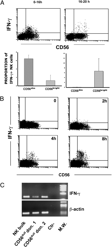
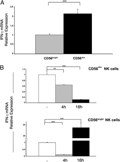
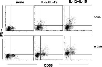
The two major functions of human natural killer (NK) cells are conventionally associated with distinct cell subsets. Thus, cytolytic activity is mostly confined to the CD56(dim)CD16(+) subset, whereas cytokine production is generally assigned to CD56(bright)CD16(+/-) cells. In this study, we reevaluated the functional capabilities of these NK subsets with regard to the production of IFN-γ at different time points after cell triggering via NKp46 and NKp30 activating receptors. Different from previous studies, cytokine production was also assessed at early intervals. We show that CD56(dim) NK cells produce IFN-γ already at 2 to 4 h, whereas no cytokine production is detected beyond 16 h. In contrast, CD56(bright) cells release IFN-γ only at late time intervals (>16 h after stimulation). The rapid IFN-γ production by CD56(dim) NK cells is in line with the presence of IFN-γ mRNA in freshly isolated cells. Rapid IFN-γ production was also induced by combinations of IL-2, IL-12, and IL-15. Our data indicate that not only cytolytic activity but also early IFN-γ production is a functional property of CD56(dim) NK cells. Thus, this subset can assure a rapid and comprehensive NK cell intervention during the early phases of innate responses.


Conflict of interest statement

The authors declare no conflict of interest.

Figures

Fig. 1.
Fig. 1.
IFN-γ production by peripheral blood CD56dim and CD56bright NK cell subset. (A) Flow cytometric analysis. PBMCs were stimulated in a redirected triggering assay (reverse antibody-dependent cell-mediated cytotoxicity) using FcγR + P815 cells and anti-NKp30/anti-NKp46 mAbs (γ isotype). GolgiPlug was added immediately at the time of stimulation until processed (Upper, 0–16 h) or after 16 h with analysis of cells after 4 h (Lower, 16–20 h; Fig. S1). CD31914 cells were gated by flow cytometric analysis and analyzed for CD56 expression and intracellular IFN-γ production. Data are representative of 12 experiments. (B) Analysis of IFN-γ production at early time points. Cell populations enriched in NK cells by depletion of CD3+, CD19+, and CD14+ cells were analyzed for CD56 expression and intracellular IFN-γ production by flow cytometry at given sequential times. Data are representative of five experiments. Numbers and bars are referred to the proportions of IFN-γ+ cells within the specific CD56dim and CD56bright subpopulations. (C) IFN-γ mRNA expression. Sorted CD56dim freshly isolated from different donors were analyzed by RT-PCR for IFN-γ mRNA expression. Polyclonal IL-2–activated NK cells were used as control. PCR products were run on a 0.8% agarose gel and visualized by ethidium bromide staining. As positive control, RT-PCR was also performed with primers specific for β-actin.
Fig. 2.
Fig. 2.
IFN-γ mRNA expression in CD56bright and CD56dim NK cell subsets. (A) IFN-γ mRNA was analyzed by real-time PCR in sorted CD56bright and CD56dim NK cells freshly isolated from six donors. Gene expression levels were normalized to GAPDH mRNA and relative quantification was performed using the ΔΔCT method. The levels of IFN-γ mRNA in CD56bright NK cells were chosen as reference and were arbitrarily normalized to 1. Data are representative of six independent experiments with six different donors (mean ± SE; ***P = 0.0001). (B) Time course: levels of IFN-γ mRNA were assessed at time 0 (white bars) or following stimulation with anti-NKp46/NKp30 mAbs for 4 h (gray bars) or 16 h (black bars). For each NK cell subset, unstimulated cells were chosen as reference and levels of IFN-γ mRNA in these cells were arbitrarily normalized to 1. All samples were analyzed in three independent experiments, processing cells obtained from two donors at a time. For each bar, the SE is indicated (**P = 0.002; ***P = 0.0001).
Fig. 3.
Fig. 3.
IFN-γ production by peripheral blood NK cell subsets following exposure to IL-2, IL-12, and IL-15. Flow cytometric analysis of peripheral blood NK cells triggered by combinations of cytokines (IL-2 plus IL-12, IL-12 plus IL-15). GolgiPlug was added immediately at the time of stimulation until processed (0–16 h) or after 16 h with analysis of cells after 4 h (16–20 h). Data are representative of four experiments.

References

    1. Caligiuri MA. Human natural killer cells. Blood. 2008;112:461–469. - PMC - PubMed
    1. Moretta A, Marcenaro E, Parolini S, Ferlazzo G, Moretta L. NK cells at the interface between innate and adaptive immunity. Cell Death Differ. 2008;15:226–233. - PubMed
    1. Moretta L, et al. Surface NK receptors and their ligands on tumor cells. Semin Immunol. 2006;18:151–158. - PubMed
    1. Bottino C, Castriconi R, Moretta L, Moretta A. Cellular ligands of activating NK receptors. Trends Immunol. 2005;26:221–226. - PubMed
    1. Moretta L, Moretta A. Killer immunoglobulin-like receptors. Curr Opin Immunol. 2004;16:626–633. - PubMed

Publication types