. 2011 Jan 11;108(2):762-7.
doi: 10.1073/pnas.1013067108. Epub 2010 Dec 27.

Biglycan recruits utrophin to the sarcolemma and counters dystrophic pathology in mdx mice

Affiliations

Biglycan recruits utrophin to the sarcolemma and counters dystrophic pathology in mdx mice

Alison R Amenta et al. Proc Natl Acad Sci U S A. .

Abstract

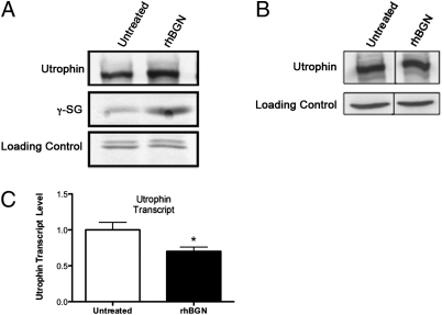
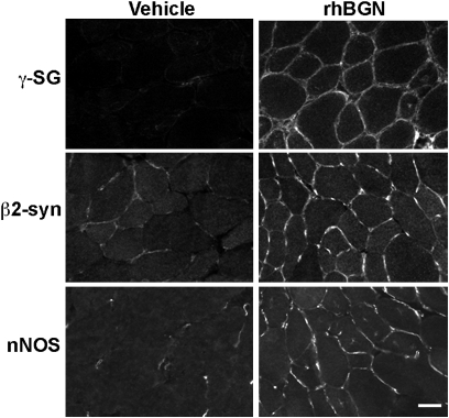
Duchenne muscular dystrophy (DMD) is caused by mutations in dystrophin and the subsequent disruption of the dystrophin-associated protein complex (DAPC). Utrophin is a dystrophin homolog expressed at high levels in developing muscle that is an attractive target for DMD therapy. Here we show that the extracellular matrix protein biglycan regulates utrophin expression in immature muscle and that recombinant human biglycan (rhBGN) increases utrophin expression in cultured myotubes. Systemically delivered rhBGN up-regulates utrophin at the sarcolemma and reduces muscle pathology in the mdx mouse model of DMD. RhBGN treatment also improves muscle function as judged by reduced susceptibility to eccentric contraction-induced injury. Utrophin is required for the rhBGN therapeutic effect. Several lines of evidence indicate that biglycan acts by recruiting utrophin protein to the muscle membrane. RhBGN is well tolerated in animals dosed for as long as 3 months. We propose that rhBGN could be a therapy for DMD.


Conflict of interest statement

Conflict of interest statement: A.R.A., B.A.M., and J.R.F. are inventors on patents owned by Brown University covering biglycan as a therapeutic for neuromuscular diseases. J.F. is a Director and shareholder in Tivorsan pharmaceuticals.

Figures

Fig. 1.
Fig. 1.
Utrophin is reduced at the sarcolemma of immature bgn−/o mice. (A) Quadriceps muscles from congenic P14 WT (Upper Panels) DJS and bgn−/o (Lower Panels) mice were harvested, sectioned, mounted on the same slides, and immunostained for dystrophin and utrophin. Utrophin expression is decreased in these developing biglycan null mice compared with WT mice, whereas dystrophin expression is not detectably altered. (Scale bar = 25 μm.) (B) Quantification of sarcolemmal utrophin expression. Images of utrophin-stained muscle sections as prepared in A were acquired and the levels of utrophin immunostaining at the perijunctional sarcolemma were measured as described in Materials and Methods. A total of 50 sarcolemmal segments from each of three animals from each genotype were analyzed. Utrophin immunoreactivity was decreased 28% in sections from bgn−/o muscle compared with WT (Bgn−/o: 0.72 ± 0.03, WT: 1.0 ± 0.04, unpaired Student t test, P < 0.0001; n = 150 sarcolemmal segments from three mice of each genotype). (C) Quantification of perijunctional sarcolemmal dystrophin. Dystrophin-stained sections were imaged and measured as in B. Dystrophin immunoreactivity was equivalent in P14 WT and bgn−/o sections (Bgn−/o: 1.01 ± 0.03, WT: 1.00 ± 0.03, unpaired Student t test, P = 0.76). (D) Quantitative real-time PCR analysis of utrophin transcripts in P14 WT and bgn−/o mice. Total RNA was extracted from quadriceps muscles from WT and bgn−/o mice and used for cDNA synthesis. Expression of utrophin mRNA was indistinguishable in WT and Bgn−/o muscles (WT: 1.0 ± 0.26, Bgn−/o: 0.99 ± 0.09, n = 3 animals from each genotype).
Fig. 2.
Fig. 2.
RhBGN treatment increases membrane-associated utrophin and γ-sarcoglycan protein in cultured myotubes. (A) Cultured bgn−/o myotubes were incubated for 8 h with either 1 nM rhBGN or vehicle as indicated. Shown are Western blots of membrane fractions probed for utrophin and γ-sarcoglycan (γ-SG). Note the increased expression of both utrophin and γ-sarcoglycan following rhBGN treatment. (B) Bgn−/o myotubes were treated as in A and whole-cell extracts were prepared. Proteins were separated by SDS/PAGE and immunoblotted for utrophin and actin (loading control). Total utrophin protein levels were similar in untreated and rhBGN treated cultures. (C) Quantitative RT-PCR analysis of untreated and rhBGN treated cultured bgn−/o myotubes. RhBGN treatment decreased utrophin transcript levels by ~30% (untreated: 1 ± 0.10; rhBGN treated: 0.7 ± 0.06; unpaired Student t test, P = 0.02; n = 6 separate experiments with three replicate flasks in each).
Fig. 3.
Fig. 3.
RhBGN treatment up-regulates utrophin at the sarcolemma of mdx mice. (A) Utrophin immunostaining of quadriceps muscles from P33 mdx littermate mice that received a single i.p. injection of either rhBGN or vehicle at P19. (Scale bar = 25 μm.) (B) Levels of immunostaining at the sarcolemma (e.g., arrows in A) of peripherally nucleated fibers. A total of 100 sarcolemmal segments from each of four animals were analyzed (two littermate pairs, one rhBGN- and one vehicle-injected animal per pair). Sarcolemmal utrophin immunoreactivity was >2.5-fold higher in sections from rhBGN- as compared with vehicle-injected animals (unpaired Student t test, P < 0.0001). (C) qRT PCR analysis of utrophin transcripts in from vehicle- or rhBGN-injected mdx mice. There was no significant difference in utrophin transcript levels in rhBGN treated mice compared with vehicle-injected controls (unpaired Student t test, P = 0.057; n = 8 vehicle- and 6 rhBGN-treated mice). (D) RhBGN treatment increases utrophin expression in muscle membrane fractions. Mdx mice from a single litter were injected at P16 and P38 (Left Pair) or P16, P38, and P63 (Right Pair) with rhBGN or vehicle. Muscles were harvested 3 wk after the last injection. (E) RhBGN treatment increases γ-sarcoglycan expression. Mdx mice were injected at 3-wk intervals starting at P14 with rhBGN or vehicle alone. Muscles were harvested at 15 wk of age and immunoblotted for γ-sarcoglycan. γ-Sarcoglycan is increased in the membrane fractions from rhBGN treated mdx mice compared with vehicle-treated animals.
Fig. 4.
Fig. 4.
RhBGN up-regulates DAPC components at the sarcolemma of mdx mice. Mdx mice were injected with rhBGN or vehicle at P18 and muscles were harvested at 32P. Sections of TA from vehicle- or rhBGN-treated animals were immunostained with antibodies to the indicated DAPC components as described in Materials and Methods. RhBGN treatment increased the expression of sarcolemmal γ-sarcoglycan, β2-syntrophin, and nNOS in mdx mice.
Fig. 5.
Fig. 5.
Systemically administered rhBGN counters dystrophic pathology in mdx mice. (A) H&E-stained sections of diaphragm from littermate mdx mice that were injected i.p. with vehicle (Upper Panels) or 100 μg rhBGN (Lower Panels) at P18 and harvested at P38. (Right Panels) Magnified view. Note the extensive areas of necrosis/regeneration and mononuclear cell infiltration in muscle from vehicle-injected as compared with rhBGN-injected mice. (Scale bars = 50 μm.) (B) RhBGN administration decreases proportion of CNFs in mdx muscle compared with vehicle-injected littermates (single injection; Materials and Methods). Percentages of CNFs were determined from H&E-stained diaphragm sections. RhBGN-treated mdx mice had ~50% fewer centrally nucleated myofibers as compared with vehicle-injected mdx mice (17.7% ± 2.8 and 9.6% ± 1.7 for vehicle- and rhBGN-injected animals, respectively; n = 13 vehicle-injected and 11 rhBGN-injected animals; unpaired Student t test, P = 0.028).
Fig. 6.
Fig. 6.
Physiological improvement of muscle in rhBGN-treated mdx mice. Mdx mice were injected at 3-wk intervals starting at P14 with either rhBGN (25 μg/injection; i.p.) or vehicle and tissue was harvested at 15 wk of age. Representative first to fifth ECCs of extensor digitorum longus (EDL) muscles from mdx mice injected with (A) vehicle, or (B) rhBGN. (C) Comparisons of ECC force drop between the first and the second, third, fourth, and fifth ECC of vehicle-treated (6.4 ± 1.2%; 12.4 ± 1.9%; 18.4 ± 2.3%; 22.2 ± 7%; n = 16) and rhBGN-treated (3.9 ± 0.3%; 7.5 ± 0.5%; 11.6 ± 0.8%; 14.9 ± 1.2%; n = 16) mdx mice, respectively. There is significant difference in the force drop between ECCs of vehicle-treated and rhBGN-treated mdx mice on the second, third, fourth, and fifth contractions (P = 0.05, 0.02, 0.01, 0.02, respectively; unpaired Student t test). (D) Average force drop between first and fifth ECC in vehicle-treated and rhBGN-treated mdx mice (22.2 ± 2.7% vs.14.9 ± 1.2%, respectively; P = 0.02; n = 16 muscles in each group; unpaired Student t test).

References

    1. Emery AEH. Duchenne Muscular Dystrophy. 2nd Ed. Oxford: Oxford University Press; 1993.
    1. Koenig M, et al. Complete cloning of the Duchenne muscular dystrophy (DMD) cDNA and preliminary genomic organization of the DMD gene in normal and affected individuals. Cell. 1987;50:509–517. - PubMed
    1. Blake DJ, Weir A, Newey SE, Davies KE. Function and genetics of dystrophin and dystrophin-related proteins in muscle. Physiol Rev. 2002;82:291–329. - PubMed
    1. Muntoni F, Torelli S, Ferlini A. Dystrophin and mutations: One gene, several proteins, multiple phenotypes. Lancet Neurol. 2003;2:731–740. - PubMed
    1. Tinsley J, et al. Expression of full-length utrophin prevents muscular dystrophy in mdx mice. Nat Med. 1998;4:1441–1444. - PubMed

Publication types

LinkOut - more resources