. 2011 Jan 11;108(2):852-7.
doi: 10.1073/pnas.1015981108. Epub 2010 Dec 27.

Trk retrograde signaling requires persistent, Pincher-directed endosomes

Affiliations

Trk retrograde signaling requires persistent, Pincher-directed endosomes

Polyxeni Philippidou et al. Proc Natl Acad Sci U S A. .

Abstract

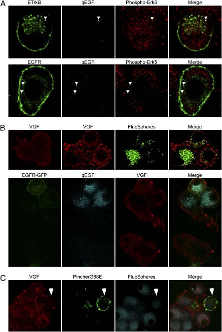
Target-derived neurotrophins use retrogradely transported Trk-signaling endosomes to promote survival and neuronal phenotype at the soma. Despite their critical role in neurotrophin signaling, the nature and molecular composition of these endosomes remain largely unknown, the result of an inability to specifically identify the retrograde signaling entity. Using EGF-bound nanoparticles and chimeric, EGF-binding TrkB receptors, we elucidate Trk-endosomal events involving their formation, processing, retrograde transport, and somal signaling in sympathetic neurons. By comparing retrograde endosomal signaling by Trk to the related but poorly neuromodulatory EGF-receptor, we find that Trk and EGF-receptor endosomes are formed and processed by distinct mechanisms. Surprisingly, Trk and EGF-receptors are both retrogradely transported to the soma in multivesicular bodies. However, only the Trk-multivesicular bodies rely on Pincher-dependent macroendocytosis and processing. Retrograde signaling through Pincher-generated Trk-multivesicular bodies is distinctively refractory to signal termination by lysosomal processing, resulting in sustained somal signaling and neuronal gene expression.


Conflict of interest statement

The authors declare no conflict of interest.

Figures

Fig. 1.
Fig. 1.
Retrogradely transported ETrkB signaling endosomes are MVBs. (A) Distal axons of chamber-cultured SCGs infected with ETrkB adenovirus were treated with qEGF for 2 h. (B) Immunocytochemistry: ETrkB (anti-EGFR, green), qEGF (red), and phospho-Erk5 (anti–P-Erk5, cyan). (Scale bar, 2 μm.) (C) EM of soma shows silver-enhanced qEGF accumulated in partially filled MVBs (Inset, arrow). (Scale bars, 500 nm.)
Fig. 2.
Fig. 2.
ETrkB, but not EGFR, retrograde transport requires both Pincher and Rac functions. (A) Chamber-cultured SCG neurons were infected with ETrkB adenovirus, the distal axons treated with qEGF525 for 6 h and neurons infected with either PincherG68E-HA or RacN17-T7 adenoviruses for 3 d, and distal axons treated for 2 h with qEGF605 (see schematic). Immunocytochemistry: qEGF525 (green), qEGF605 (cyan), PincherG68E (anti-HA, red, Top and Middle), and RacN17 (anti-T7, red, Bottom). (B) Distal axons of chamber-cultured SCGs coinfected with adenoviruses expressing ETrkB and either RacV12-GFP (Upper) or Pincher-HA (Lower) were treated with qEGF for 2 h. Immunocytochemistry: ETrkB (anti-EGFR, red), qEGF (cyan), RacV12-GFP (GFP, green, Upper) and Pincher-HA (anti-Pincher, green, lower). Arrowheads point to receptor/qEGF complexes. (C) Distal axons of chamber-cultured SCGs coinfected with adenoviruses expressing PincherG68E-HA or either ETrkB (Upper) or EGFR (Lower) were treated for 48 h with fluoSpheres and then with qEGF for 2 h. ETrkB (anti-EGFR, red, Upper), EGFR (anti-EGFR, red, Lower), PincherG68E (anti-Pincher, cyan), fluoSpheres (green), and qEGF (purple). (Scale bars, 2 μm.)
Fig 3.
Fig 3.
Retrogradely transported ETrkB and EGFR endosomes are differentially processed by Rab5 and Rab7. Chamber-cultured SCG neurons were infected with either ETrkB (B, Upper) or EGFR (B, Lower) adenovirus alone, or also infected with either Rab5-GFP (A, Upper) or Rab7-GFP (A, Lower) adenovirus. After 2 d, the distal axons were treated with qEGF for 2 h (A) or 4 h (B). ETrkB (anti-EGFR, red, Upper), EGFR (anti-EGFR, red, Lower), qEGF (cyan), Rab5-GFP (GFP, green, A, Upper), Rab7 (GFP, green, A, Lower), and cathepsin (anti-cathepsin, green, B). Arrowheads point to receptor/qEGF complexes. (Scale bars, 2 μm.)
Fig. 4.
Fig. 4.
ETrkB-MVBs retain Rab5. Chamber-cultured SCG neurons were (A) infected 48 h with an adenovirus expressing either ETrkB (Left) or EGFR (Right), the distal axons treated with qEGF for 2 h, and processed for silver-enhanced EM as in Fig. 2C (Scale bar, 200 nm), or (B) coinfected 48 h with ETrkB and Rab5-GFP adenoviruses and distal axons treated 2 h with EGF-nG. (Scale bar, 50 nm.) Somata were processed for EM, with pre-embed gold-enhanced and postembedded immuno-gold–labeling for GFP. Two double-labeled MVBs are shown.
Fig. 5.
Fig. 5.
Retrogradely transported endosomes containing ETrkB, but not EGFR, mediated sustained Erk signaling and vgf gene induction. Chamber-cultured SCG neurons were: (A) infected 48 h with either ETrkB (Upper) or EGFR (Lower) adenovirus, and the distal axon treated 2 h with qEGF. ETrkB (anti-EGFR, green, Upper), EGFR (anti-EGFR, green, Lower), qEGF (cyan), and phospho-Erk5 (anti-phospho-Erk5, red); arrowheads point to receptor/qEGF complexes; (B) untreated (Upper Left), treated at distal axons of uninfected (Upper), or EGFR-GFP adenovirus-infected (Lower) neurons with fluoSpheres and NGF (Upper Right) or qEGF alone (Lower) for 10 h, VGF (anti-VGF, red), fluoSpheres (green, Upper), EGFR-GFP (GFP, green, Lower), and qEGF (cyan, Lower); (C) NGF, starved for 48 h using anti-NGF and infected at distal axons with a PincherG68E-HA HSV and then treated for 10 h with fluoSpheres and NGF. VGF (anti-VGF, red), PincherG68E (anti-HA, green), and fluoSpheres (cyan). Arrowheads points to PincherG68E-expressing cell.

References

    1. Levi-Montalcini R. The nerve growth factor 35 years later. Science. 1987;237:1154–1162. - PubMed
    1. Zweifel LS, Kuruvilla R, Ginty DD. Functions and mechanisms of retrograde neurotrophin signalling. Nat Rev Neurosci. 2005;6:615–625. - PubMed
    1. Halegoua S, Armstrong RC, Kremer NE. Dissecting the mode of action of a neuronal growth factor. Curr Top Microbiol Immunol. 1991;165:119–170. - PubMed
    1. Beattie EC, et al. A signaling endosome hypothesis to explain NGF actions: Potential implications for neurodegeneration. Cold Spring Harb Symp Quant Biol. 1996;61:389–406. - PubMed
    1. Valdez G, et al. Trk-signaling endosomes are generated by Rac-dependent macroendocytosis. Proc Natl Acad Sci USA. 2007;104:12270–12275. - PMC - PubMed

Publication types