. 2011 Jan 11;108(2):592-7.
doi: 10.1073/pnas.1013643108. Epub 2010 Dec 27.

Structural studies of ion permeation and Ca2+ blockage of a bacterial channel mimicking the cyclic nucleotide-gated channel pore

Affiliations

Structural studies of ion permeation and Ca2+ blockage of a bacterial channel mimicking the cyclic nucleotide-gated channel pore

Mehabaw G Derebe et al. Proc Natl Acad Sci U S A. .

Abstract

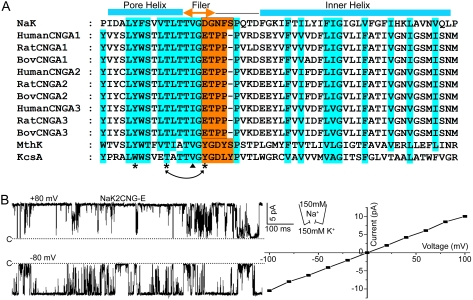
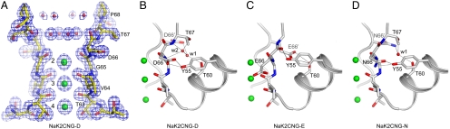
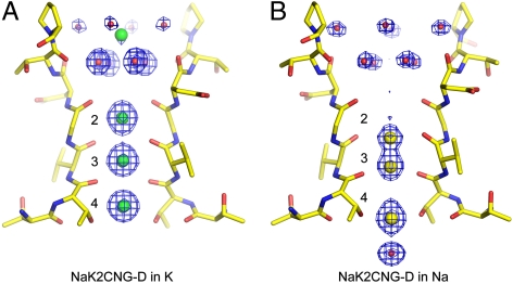
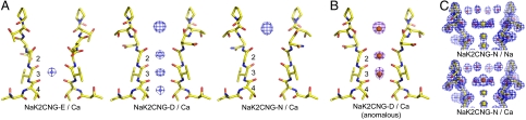
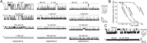
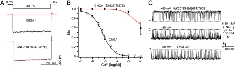
Cyclic nucleotide-gated (CNG) channels play an essential role in the visual and olfactory sensory systems and are ubiquitous in eukaryotes. Details of their underlying ion selectivity properties are still not fully understood and are a matter of debate in the absence of high-resolution structures. To reveal the structural mechanism of ion selectivity in CNG channels, particularly their Ca(2+) blockage property, we engineered a set of mimics of CNG channel pores for both structural and functional analysis. The mimics faithfully represent the CNG channels they are modeled after, permeate Na(+) and K(+) equally well, and exhibit the same Ca(2+) blockage and permeation properties. Their high-resolution structures reveal a hitherto unseen selectivity filter architecture comprising three contiguous ion binding sites in which Na(+) and K(+) bind with different ion-ligand geometries. Our structural analysis reveals that the conserved acidic residue in the filter is essential for Ca(2+) binding but not through direct ion chelation as in the currently accepted view. Furthermore, structural insight from our CNG mimics allows us to pinpoint equivalent interactions in CNG channels through structure-based mutagenesis that have previously not been predicted using NaK or K(+) channel models.


Conflict of interest statement

The authors declare no conflict of interest.

Figures

Fig. 1.
Fig. 1.
(A) Partial sequence alignment between NaK, K+ channels (MthK and KcsA) and human, rat, and bovine CNG channel alpha subunits (A1–A3). Semiconserved residues are shaded in cyan. Residues in NaK replaced by corresponding CNG channel residues are shaded in orange. Secondary structure assignment is based on the NaKNΔ19 structures (PDB ID 3E86). Asterisks mark the positions where mutagenesis was performed on bovine CNGA1 based on the NaK2CNG-E structure and arrows indicate the residues for swap mutations. (B) Single channel traces of NaK2CNG-E at ± 80 mV and its I-V curve. Currents were recorded using giant liposome patch clamping with 150 mM NaCl and 150 mM KCl in the pipette and bath solutions, respectively. Dotted lines mark the zero current level.
Fig. 2.
Fig. 2.
Selectivity filter structures of CNG-mimicking NaK mutants. (A) Selectivity filter structure of NaK2CNG-D in complex with K+ ions. The 2Fo-Fc electron density map (1.62 Å) is contoured at 2.0σ (blue mesh). Red spheres represent the two layers of water molecules within the external funnel. Three ion binding sites are labeled 2–4 from top to bottom. (BD) Protein packing around the selectivity filters of (B) NaK2CNG-D, (C) NaK2CNG-E, and (D) NaK2CNG-N. Red spheres represent water molecules that mediate the hydrogen bonding network. Residue 66 from the neighboring subunit is labeled as D66′, E66′, and N66′, respectively. All K+ ions in the filter are drawn as green spheres.
Fig. 3.
Fig. 3.
K+ and Na+ binding in the selectivity filter of NaK2CNG-D (A) Fo-Fc ion omit map focusing on the selectivity filter of the NaK2CNG-D-K+ complex (1.62 Å) contoured at 4σ. (B) Fo-Fc ion omit maps focusing on the selectivity filter of the NaK2CNG-D-Na+ complex (1.58 Å) contoured at 4σ. Water molecules within the external funnel (red spheres) were also omitted in the map calculation. K+ and Na+ ions are represented by green and yellow spheres, respectively.
Fig. 4.
Fig. 4.
Ca2+ binding in the selectivity filter of NaK mutants. (A) FNa/Ca-soak-FNa-soak difference Fourier maps between mutant crystals soaked in stabilization solutions containing 100 mM NaCl (Na-soak) and in solutions containing both 100 mM NaCl and 25 mM CaCl2 (Na/Ca-soak). Maps are contoured at 8σ at a resolution of 2.0 Å for NaK2CNG-E (Left) and 1.8 Å for NaK2CNG-D (Center) and NaK2CNG-N (Right). (B) Anomalous difference Fourier map (purple mesh, at 1.9 Å and contoured at 5σ) of a Na/Ca-soaked NaK2CNG-D crystal indicates the positions of bound Ca2+ ions (orange spheres). (C) 2Fo-Fc maps of Na-soaked (Upper, 1.62 Å) and Na/Ca-soaked (Upper, 1.63 Å) NaK2CNG-N crystals reveal the hydration of an externally bound Na+ (yellow sphere) or Ca2+ ion (orange sphere) by the inner layer of water molecules within the funnel. External Ca2+ in NaK2CNG-D is bound in the same hydrated form.
Fig. 5.
Fig. 5.
Ca2+ blockage and permeation in NaK mutants. (A) Single channel traces of NaK2CNG-E (Left traces), NaK2CNG-D (Center traces), and NaK2CNG-N (Right traces) recorded at -80 mV in the presence of various concentrations of external Ca2+. Both pipette and bath solutions contain symmetrical 150 mM KCl. Additionally, 30 μM tetrapentyl ammonium (TPeA) was added to the bath solution to ensure that recorded currents are from mutant channels oriented with their external side facing the bath solution. (B) Plot of the fraction of unblocked single channel currents recorded at -80 mV as a function of external Ca2+ concentrations. Data points are mean ± SEM from five measurements and are fitted to the Hill equation with Ki of 58.3 ± 6.3 μM and Hill coefficient n = 0.98 ± 0.1 for NaK2CNG-E, and Ki of 6.3 ± 0.62 μM and n = 1.22 ± 0.1 for NaK2CNG-D. (C) Single channel traces of NaK2CNG-E recorded at -80 mV with 150 mM KCl in the pipette and 100 mM CaCl2 in the bath solution. The single channel in the recordings had its intracellular side facing the bath solution and was blocked by addition of 30 μM TPeA in bath solution (bottom trace).
Fig. 6.
Fig. 6.
Mutagenesis of bovine CNGA1 channel based on the structure of NaK2CNG-E. (A) Macroscopic currents of wild-type bovine retinal CNGA1 (Upper) and its E363T/T357E swapped mutation (Lower) recorded at -80 mV in the presence (red traces) and absence (black traces) of 100 μM extracellular Ca2+. Channels were activated by 1 mM cGMP in the pipette solution. (B) Plot of the fraction of unblocked inward currents (I/Io) at -80 mV as a function of external Ca2+ concentrations. Data points are mean ± SEM from five measurements and are fitted to Langmuir functions with Ki of 7.1 μM for wild-type CNGA1. (C) Single channel traces of NaK2CNG-E (E66T/T60E) swapped mutation recorded at ± 80 mV with 150 mM NaCl in the pipette and 150 mM KCl in the bath solution (top two traces). Addition of 1 mM Ca2+ at extracellular side (bath solution) has no effect on single channel conductance at -80 mV (bottom trace).

Comment in

References

    1. Yau KW, Baylor DA. Cyclic GMP-activated conductance of retinal photoreceptor cells. Annu Rev Neurosci. 1989;12:289–327. - PubMed
    1. Kaupp UB, Seifert R. Cyclic nucleotide-gated ion channels. Physiol Rev. 2002;82:769–824. - PubMed
    1. Matulef K, Zagotta WN. Cyclic nucleotide-gated ion channels. Annu Rev Cell Dev Biol. 2003;19:23–44. - PubMed
    1. Zagotta WN, Siegelbaum SA. Structure and function of cyclic nucleotide-gated channels. Annu Rev Neurosci. 1996;19:235–263. - PubMed
    1. Baumann A, Frings S, Godde M, Seifert R, Kaupp UB. Primary structure and functional expression of a Drosophila cyclic nucleotide-gated channel present in eyes and antennae. EMBO J. 1994;13:5040–5050. - PMC - PubMed

Publication types

LinkOut - more resources